. 2017 Jan;103(1):117-133.
doi: 10.1111/mmi.13546. Epub 2016 Oct 27.

The Paracoccus denitrificans NarK-like nitrate and nitrite transporters-probing nitrate uptake and nitrate/nitrite exchange mechanisms

Affiliations

The Paracoccus denitrificans NarK-like nitrate and nitrite transporters-probing nitrate uptake and nitrate/nitrite exchange mechanisms

Alan D Goddard et al. Mol Microbiol. 2017 Jan.

Abstract

Nitrate and nitrite transport across biological membranes is often facilitated by protein transporters that are members of the major facilitator superfamily. Paracoccus denitrificans contains an unusual arrangement whereby two of these transporters, NarK1 and NarK2, are fused into a single protein, NarK, which delivers nitrate to the respiratory nitrate reductase and transfers the product, nitrite, to the periplasm. Our complementation studies, using a mutant lacking the nitrate/proton symporter NasA from the assimilatory nitrate reductase pathway, support that NarK1 functions as a nitrate/proton symporter while NarK2 is a nitrate/nitrite antiporter. Through the same experimental system, we find that Escherichia coli NarK and NarU can complement deletions in both narK and nasA in P. denitrificans, suggesting that, while these proteins are most likely nitrate/nitrite antiporters, they can also act in the net uptake of nitrate. Finally, we argue that primary sequence analysis and structural modelling do not readily explain why NasA, NarK1 and NarK2, as well as other transporters from this protein family, have such different functions, ranging from net nitrate uptake to nitrate/nitrite exchange.


Figures

Figure 1
Figure 1
Schematic representation of the nitrate/nitrite transporters of P. denitrificans, discussed in this study, along with some of the proteins interacting with nitrate or nitrite on either side of the inner membrane. In P. denitrificans, NarK1 and NarK2 are a single protein fusion called NarK. Proteins involved in the assimilation pathway are shown in white while proteins involved in respiration are shown in black. Nitrate respiration occurs under anaerobic conditions whatever the nitrogen source, whereas assimilatory proteins are expressed aerobically or anaerobically only when preferred nitrogen sources, such as ammonium, are absent. Uncertainties about the roles of NasA and NarK1/NarK2 are discussed in this study. Nar, and not Nap, is the default enzyme for respiration.
Figure 2
Figure 2
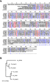
Bioinformatics analysis of nitrate/nitrite transporters. A. Alignment of P. denitrificans NasA (Pd_NasA), NarK1 (Pd_NarK1) and NarK2 (Pd_NarK2), E. coli NarK (Ec_NarK) and NarU (Ec_NarU), and A. nidulans CRNA* (An_CRNA* lacks residues 221–310 of the WT protein sequence; the extended predicted intracellular loop of WT An_CRNA (residues 221–310) was removed because it compromises the alignment) was performed using Multalin (Corpet, 1988). An_CRNA* is included to give a comparison with a eukaryotic NarK‐like protein. Residues which are conserved in all proteins are shown in red whereas residues which are conserved in half or more of the proteins are in blue. B. Phylogenetic tree of the same nitrate/nitrite transporter as shown in (A). In this case, the WT sequence of An_CRNA was used. The tree was constructed using Multalin (Corpet, 1988). The scale bar represents 10 point accepted mutations (PAM).
Figure 3
Figure 3
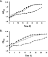
Complementation of ΔnasA and ΔnasA ΔnasH P. denitrificans strains by P. denitrificans NarK‐like proteins. P. denitrificans (A) ΔnasA or (B) ΔnasA ΔnasH were grown aerobically in the presence of nitrate as a sole nitrogen source, while harboring pEG276‐derived plasmids expressing full‐length P. denitrificans NarK (⋄), P. denitrificans NarK1 (Δ), P. denitrificans NarK2 (○), P. denitrificans NasA (+) or empty vector (*). WT Pd1222 is indicated by (□). Extracellular nitrite accumulation was also determined in the same strains ((C) for ΔnasA and (D) for ΔnasA ΔnasH). Three biological replicates were performed for each experiment and representative data is shown.
Figure 4
Figure 4
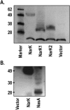
Analysis of the expression of the NarK and NasA proteins in the ΔnasA P. denitrificans strain. A. NarK full length, NarK1 or NarK2 and (B) NasA were expressed from equivalent constructs with a C‐terminal hexahistidine‐tag. Extracts from P. denitrificans ΔnasA strains expressing the relevant protein were analysed by SDS‐PAGE, followed by Western blot analysis using an anti‐pentahistidine antibody (Qiagen). NarK1, NarK2 and NasA migrate faster than predicted based on their molecular weight; this is common for integral membrane proteins and it is consistent with previous studies (Goddard et al., 2008). The protein band detected in all lanes of (A), migrating below the 28 kDa protein standard, is due to non‐specific binding of the antibody. The same expression pattern was observed in a ΔnasA ΔnasH strain.
Figure 5
Figure 5
Complementation of ΔnasA and ΔnasA ΔnasH P. denitrificans strains by E. coli NarK‐like proteins. P. denitrificans (A) ΔnasA or (B) ΔnasA ΔnasH were grown aerobically in the presence of nitrate as a sole nitrogen source, while harboring pEG276‐derived plasmids expressing E. coli NarK (black ○), E. coli NarU (light grey ⋄) and the empty expression vector (dark grey Δ). The results shown are the average of three biological replicates.
Figure 6
Figure 6
Schematic representation of P. denitrificans NarK demonstrating the location of important residues. The 24 transmembrane helices and connecting loops of NarK, as predicted by the TMHMM program, are illustrated and the relative positions of residues mutated in this study are marked. White circles represent non‐essential amino acids and black circles represent residues that abolish the growth of a ΔnarK strain complemented with the appropriate plasmid; mutations in the essential arginines have been described previously (Goddard et al., 2008). M442, the start codon of NarK2, is indicated with a grey circle.
Figure 7
Figure 7
Analysis of the expression of NarK1 and NarK2 proline variants. For each of the NarK1 or NarK2 proline mutations which affected the function of these domains, the proline variant was expressed from a construct with a C‐terminal hexahistidine‐tag. Extracts from P. denitrificans ΔnarK strains expressing the relevant variant were analysed by SDS‐PAGE followed by Western blot analysis using an anti‐pentahistidine antibody (Qiagen). NarK1 and NarK2 migrate faster than predicted based on their molecular weight; this is common for integral membrane proteins and it is consistent with previous studies (Goddard et al., 2008). The protein band detected in all lanes, migrating below the 28 kDa protein standard, is due to non‐specific binding of the antibody.
Figure 8
Figure 8
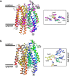
Overlays of the homology models of P. denitrificans NarK‐like transporters on the crystal structures of the E. coli nitrate transporters. A. Overlay of the homology models of P. denitrificans NarK1 and NasA on the crystal structure of E. coli NarK. The crystal structure of E. coli NarK (PDB: 4U4V) is shown in cartoon and is coloured from the N‐terminus (blue) to the C‐terminus (red) while the homology models of NarK1 and NasA are shown in grey and magenta respectively. The main residues involved in nitrate binding are strictly conserved in NarK1 and NasA and overlay extremely well onto the equivalent side chains of E. coli NarK (active‐site inset). B. Overlay of the homology model of P. denitrificans NarK2 on the crystal structure of E. coli NarU. The crystal structure of E. coli NarU (PDB: 4IU8) is shown in cartoon and is coloured from the N‐terminus (blue) to the C‐terminus (red) while the homology model of NarK2 is shown in grey. The main residues involved in nitrate binding are strictly conserved in NarK2 and overlay extremely well onto the equivalent side chains of E. coli NarU (active‐site inset).

Similar articles

Cited by

References

    1. Abramson, J. , Smirnova, I. , Kasho, V. , Verner, G. , Kaback, H.R. , and Iwata, S. (2003) Structure and mechanism of the lactose permease of Escherichia coli . Science 301: 610–615. - PubMed
    1. Abramson, J. , Iwata, S. , and Kaback, H.R. (2004) Lactose permease as a paradigm for membrane transport proteins. Mol Membr Biol 21: 227–236. - PubMed
    1. Alefounder, P.R. , Mccarthy, J.E.G. , and Ferguson, S.J. (1981) The basis of the control of nitrate reduction by oxygen in Paracoccus dentrificans . FEMS Microbiol Lett 12: 321–326.
    1. Boogerd, F.C. , Vanverseveld, H.W. , and Stouthamer, A.H. (1983) Dissimilatory nitrate uptake in Paracoccus denitrificans via a Δ˜μH+‐dependent system and a nitrate‐nitrite antiport system. Biochim Biophys Acta 723: 415–427.
    1. Borrero‐de Acuna, J.M. , Rohde, M. , Wissing, J. , Jansch, L. , Schobert, M. , Molinari, G. , et al (2016) Protein network of the Pseudomonas aeruginosa denitrification apparatus. J Bacteriol 198: 1401–1413. - PMC - PubMed

MeSH terms