Review
. 2016 Nov/Dec;51(6):513-527.
doi: 10.1080/10409238.2016.1237934. Epub 2016 Oct 4.

Decoding P4-ATPase substrate interactions

Affiliations
Review

Decoding P4-ATPase substrate interactions

Bartholomew P Roland et al. Crit Rev Biochem Mol Biol. 2016 Nov/Dec.

Abstract

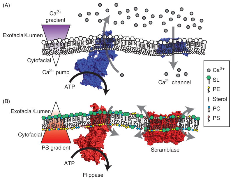
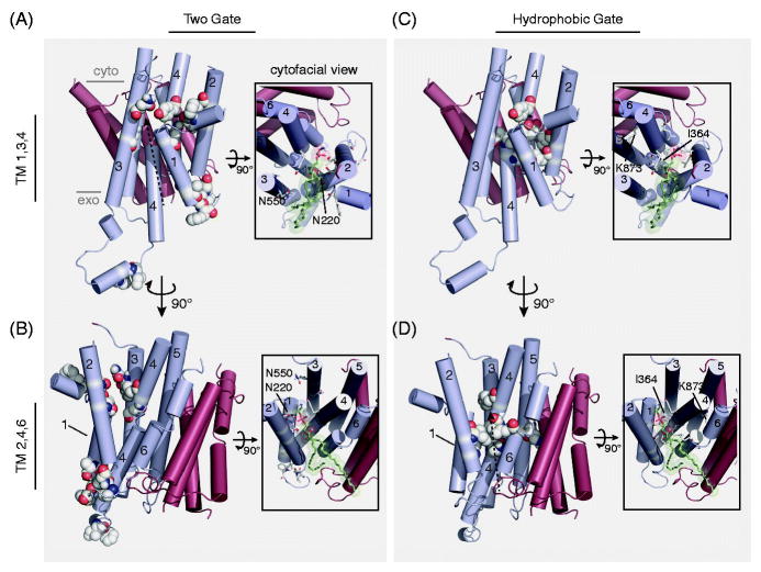
Cellular membranes display a diversity of functions that are conferred by the unique composition and organization of their proteins and lipids. One important aspect of lipid organization is the asymmetric distribution of phospholipids (PLs) across the plasma membrane. The unequal distribution of key PLs between the cytofacial and exofacial leaflets of the bilayer creates physical surface tension that can be used to bend the membrane; and like Ca2+, a chemical gradient that can be used to transduce biochemical signals. PL flippases in the type IV P-type ATPase (P4-ATPase) family are the principle transporters used to set and repair this PL gradient and the asymmetric organization of these membranes are encoded by the substrate specificity of these enzymes. Thus, understanding the mechanisms of P4-ATPase substrate specificity will help reveal their role in membrane organization and cell biology. Further, decoding the structural determinants of substrate specificity provides investigators the opportunity to mutationally tune this specificity to explore the role of particular PL substrates in P4-ATPase cellular functions. This work reviews the role of P4-ATPases in membrane biology, presents our current understanding of P4-ATPase substrate specificity, and discusses how these fundamental aspects of P4-ATPase enzymology may be used to enhance our knowledge of cellular membrane biology.

Keywords: P4-ATPase; membrane asymmetry; membrane biology; phospholipid flippase; phospholipid transport; protein engineering.


Conflict of interest statement

Declaration of Interest This work was supported by the National Institutes of Health (R01-GM107978 TRG; F32-GM116310 BPR).

Figures

Figure 1
Figure 1. A comparison of Ca2+ and phospholipid gradients
A) Cellular Ca2+ is tightly regulated. At rest, Ca2+ is actively transported into the lumen of the ER and across the PM to the extracellular fluid by Ca2+ pumps, creating a polarized gradient of Ca2+ ions across the membrane. When activated, Ca2+ channels open to harness this chemical potential and transduce cellular signals by rapidly increasing the cytosolic Ca2+ concentration. B) Similar to the establishment of the Ca2+ gradient, lipid flippases (P4-ATPases) will selectively transport glycerophospholipid species from the lumenal or exofacial aspect to the cytofacial leaflet, creating an asymmetric distribution of phospholipids across the membrane. In this example, PS is retained on the cytofacial leaflet of the membrane. Once this PS gradient is set, scramblase activation will non-selectively translocate lipid species between the leaflets of the bilayer to transduce cellular signals. In legend: calcium (Ca2+), sphingolipid (SL), phosphatidylethanolamine (PE), cholesterol in mammals or ergosterol in yeast (sterol), phosphatidylcholine (PC), phosphatidylserine (PS). Ca2+ pump: sarco/endoplasmic reticulum Ca2+ ATPase (PDB: 3W5D). Scramblase: TMEM16F (PDB: 4WIT). Ca2+ channel: voltage-gated Ca2+ channel (PDB: 4MS2). Flippase: Dnf1 homology model (Roland & Graham, 2016). Color figures can be found online.
Figure 2
Figure 2. P4-ATPase domain organization and proposed 2-gate mechanism of phospholipid translocation
Most P4-ATPases are composed of a catalytic α subunit and a noncatalytic β subunit (Cdc50). Movements of the nucleotide binding (N), phosphorylation (P), actuator (A) and membrane domain helices during catalysis are modeled after SERCA1 structures trapped in the indicated conformational states. Clusters of residues that determine substrate specificity in TM1-4 labeled as the entry and exit gates. A substrate phospholipid is shown in green using the “Credit Card” model of transport to the cytosolic leaflet. Color figures can be found online.
Figure 3
Figure 3. The anatomy of a P4-ATPase phospholipid substrate
A dioleoyl phosphatidylserine (DOPS) is used to depict the three key molecular positions that P4-ATPases use to discriminate their substrate: i) the headgroup, ii) the glycerol backbone, and iii) the acyl chain linkages. Presented at each PL molecular position are different modifications that have been tested as P4-ATPase substrates from a variety of enzymes, with positive and negative observations indicated. Selected citations – headgroup: (Seigneuret and Devaux, 1984, Zachowski et al., 1985, Zachowski et al., 1986, Drummond and Daleke, 1995, Paterson et al., 2006, Smriti et al., 2007); backbone: (Morrot et al., 1989, Pomorski et al., 2003, Smriti et al., 2007); acyl chains: (Morrot et al., 1989, Fellmann et al., 1993, Fellmann et al., 2000, Paterson et al., 2006).
Figure 4
Figure 4. Working toward a P4-ATPase barcode
A ClustalO alignment of yeast and human P4-ATPases: Uniprot accession numbers S.cerevisiae Dnf1 P32660, S.cerevisiae Dnf2 Q12675, S.cerevisiae Drs2 P39524, H.sapiens ATP8A1 Q9Y2Q0, H.sapiens ATP8A2 Q9NTI2, H.sapiens ATP8B1 O43520, H.sapiens ATP8B2 P98198, H.sapiens ATP10A O60312, H.sapiens ATP11A P98196, H.sapiens APT11C Q8NB49. Shown are alignment fragments highlighting TMs 1,2,3 and 4. The alignments are divided into two clades: the PC transporters and PS transporters. Positions that have been demonstrated to change substrate selectivity are color-coded, with specific gain-of-function mutations indicated with arrows. For example, G230,A231 in Dnf1 prevent PS transport in WT Dnf1, but mutations to the Drs2 residues QQ allows Dnf1 to transport PS. Conversely, the Drs2 QQ → GA mutation disrupts PS transport, but this mutant retains the ability to flip PE. The conserved P-type ATPase TM4 proline is indicated in Bold as a point of reference. Color figures can be found online.
Figure 5
Figure 5. A comparison of putative P4-ATPase substrate pathways
A,B) The “Two-gate” hypothesis of substrate passage proposes the recognition of PL headgroup at an exofacial/lumenal entry gate (G230, A231, I234, F235, P240, G241, F587, I580) and transport to the cytofacial exit gate (F213, N220, T254, D258, N550, N556, Y618) (numbers from Dnf1). C,D) The “Hydrophobic gate” hypothesis of substrate passage contends that a cluster of hydrophobic residues forming an internal channel to alternate a water-solvated pathway for substrate passage (F88, L112, I115, I362, I364, L367, V908), influenced by the participation of K873 (numbers from ATP8A2). These two hypotheses are not mutually exclusive, and are illustrated by identical dashed-arrows proposing substrate trajectory through the enzyme models. The principle dispute between the two hypotheses is the orientation of the PL substrate during translocation: are the acyl chains directed toward a cleft formed by TM1,3,4 (A,C); or TM2,4,6 (B,D)? Each pathway is depicted using a homology model derived from SERCA (PDB: 3W5D) with a PE molecule docked at the cytofacial aspect in the TM1,3,4 (A,C) and TM2,4,6 (B,D) orientation (Roland and Graham, 2016). Only the TM domain is depicted, with alpha helices shown in cylinders. TM segments 1–6 are colored cyan, while TMs 7–10 are red. Major images: lateral view of the TM domain, substrate-selective residues are colored white/by elements and shown in spheres. Image insets: cytofacial view of the TM domain, substrate-selective residues are colored white/by elements and shown in sticks, docked PE model is colored green/by element and shown in sticks with transparent spheres to illustrate space accommodations. Select residues are numbered and indicated (Dnf1 – Two-gate model; ATP8A2 – hydrophobic gate model). Color figures can be found online.

References

    1. Alder-Baerens N, Lisman Q, Luong L, Pomorski T, Holthuis JC. Loss of P4 ATPases Drs2p and Dnf3p disrupts aminophospholipid transport and asymmetry in yeast post-Golgi secretory vesicles. Mol Biol Cell. 2006;17:1632–42. - PMC - PubMed
    1. Amara A, Mercer J. Viral apoptotic mimicry. Nat Rev Microbiol. 2015;13:461–9. - PMC - PubMed
    1. Andersen JP, Vestergaard AL, Mikkelsen SA, Mogensen LS, Chalat M, Molday RS. P4-ATPases as Phospholipid Flippases-Structure, Function, and Enigmas. Front Physiol. 2016;7:275. - PMC - PubMed
    1. Arashiki N, Takakuwa Y, Mohandas N, Hale J, Yoshida K, Ogura H, Utsugisawa T, Ohga S, Miyano S, Ogawa S, Kojima S, Kanno H. ATP11C is a major flippase in human erythrocytes and its defect causes congenital hemolytic anemia. Haematologica. 2016;101:559–65. - PMC - PubMed
    1. Axelsen KB, Palmgren MG. Evolution of substrate specificities in the P-type ATPase superfamily. J Mol Evol. 1998;46:84–101. - PubMed

Substances

LinkOut - more resources