Expression and characterization of transmembrane and coiled-coil domain family 3
- PMID: 27697108
- PMCID: PMC5346324
- DOI: 10.5483/bmbrep.2016.49.11.151
Expression and characterization of transmembrane and coiled-coil domain family 3
Abstract
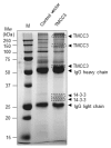
Transmembrane and coiled-coil domain family 3 (TMCC3) has been reported to be expressed in the human brain; however, its function is still unknown. Here, we found that expression of TMCC3 is higher in human whole brain, testis and spinal cord compared to other human tissues. TMCC3 was expressed in mouse developing hind brain, lung, kidney and somites, with strongest expression in the mesenchyme of developing tongue. By expression of recombinant TMCC3 and its deletion mutants, we found that TMCC3 proteins self-assemble to oligomerize. Immunostaining and confocal microscopy data revealed that TMCC3 proteins are localized in endoplasmic reticulum through transmembrane domains. Based on immunoprecipitation and mass spectroscopy data, TMCC3 proteins associate with TMCC3 and 14-3-3 proteins. This supports the idea that TMCC3 proteins form oligomers and that 14-3-3 may be involved in the function of TMCC3. Taken together, these results may be useful for better understanding of uncharacterized function of TMCC3. [BMB Reports 2016; 49(11): 629-634].
Figures


Similar articles
-
Transmembrane and coiled-coil domain family 3 (TMCC3) regulates breast cancer stem cell and AKT activation.Oncogene. 2021 Apr;40(16):2858-2871. doi: 10.1038/s41388-021-01729-1. Epub 2021 Mar 19. Oncogene. 2021. PMID: 33742122 Free PMC article.
-
TMCC3 localizes at the three-way junctions for the proper tubular network of the endoplasmic reticulum.Biochem J. 2019 Nov 15;476(21):3241-3260. doi: 10.1042/BCJ20190359. Biochem J. 2019. PMID: 31696206
-
The 14-3-3γ isoform binds to and regulates the localization of endoplasmic reticulum (ER) membrane protein TMCC3 for the reticular network of the ER.J Biol Chem. 2023 Feb;299(2):102813. doi: 10.1016/j.jbc.2022.102813. Epub 2022 Dec 20. J Biol Chem. 2023. PMID: 36549645 Free PMC article.
-
Transmembrane and coiled-coil domain family 3 gene is a novel target of hepatic peroxisome proliferator-activated receptor γ in fatty liver disease.Mol Cell Endocrinol. 2024 Dec 1;594:112379. doi: 10.1016/j.mce.2024.112379. Epub 2024 Sep 24. Mol Cell Endocrinol. 2024. PMID: 39326649
-
EHD1--an EH-domain-containing protein with a specific expression pattern.Genomics. 1999 Jul 1;59(1):66-76. doi: 10.1006/geno.1999.5800. Genomics. 1999. PMID: 10395801
Cited by
-
FAM81A is a postsynaptic protein that regulates the condensation of postsynaptic proteins via liquid-liquid phase separation.PLoS Biol. 2024 Mar 7;22(3):e3002006. doi: 10.1371/journal.pbio.3002006. eCollection 2024 Mar. PLoS Biol. 2024. PMID: 38452102 Free PMC article.
-
Transmembrane and coiled-coil domain family 3 (TMCC3) regulates breast cancer stem cell and AKT activation.Oncogene. 2021 Apr;40(16):2858-2871. doi: 10.1038/s41388-021-01729-1. Epub 2021 Mar 19. Oncogene. 2021. PMID: 33742122 Free PMC article.
-
Insights into the molecular underlying mechanisms and therapeutic potential of endoplasmic reticulum stress in sensorineural hearing loss.Front Mol Neurosci. 2024 Dec 18;17:1443401. doi: 10.3389/fnmol.2024.1443401. eCollection 2024. Front Mol Neurosci. 2024. PMID: 39744539 Free PMC article. Review.
References
MeSH terms
Substances
LinkOut - more resources
Full Text Sources
Other Literature Sources
Molecular Biology Databases

