. 2016 Oct 17;213(11):2365-2382.
doi: 10.1084/jem.20160027. Epub 2016 Oct 3.

Complement pathway amplifies caspase-11-dependent cell death and endotoxin-induced sepsis severity

Affiliations

Complement pathway amplifies caspase-11-dependent cell death and endotoxin-induced sepsis severity

Brooke A Napier et al. J Exp Med. .

Abstract

Cell death and release of proinflammatory mediators contribute to mortality during sepsis. Specifically, caspase-11-dependent cell death contributes to pathology and decreases in survival time in sepsis models. Priming of the host cell, through TLR4 and interferon receptors, induces caspase-11 expression, and cytosolic LPS directly stimulates caspase-11 activation, promoting the release of proinflammatory cytokines through pyroptosis and caspase-1 activation. Using a CRISPR-Cas9-mediated genome-wide screen, we identified novel mediators of caspase-11-dependent cell death. We found a complement-related peptidase, carboxypeptidase B1 (Cpb1), to be required for caspase-11 gene expression and subsequent caspase-11-dependent cell death. Cpb1 modifies a cleavage product of C3, which binds to and activates C3aR, and then modulates innate immune signaling. We find the Cpb1-C3-C3aR pathway induces caspase-11 expression through amplification of MAPK activity downstream of TLR4 and Ifnar activation, and mediates severity of LPS-induced sepsis (endotoxemia) and disease outcome in mice. We show C3aR is required for up-regulation of caspase-11 orthologues, caspase-4 and -5, in primary human macrophages during inflammation and that c3aR1 and caspase-5 transcripts are highly expressed in patients with severe sepsis; thus, suggesting that these pathways are important in human sepsis. Our results highlight a novel role for complement and the Cpb1-C3-C3aR pathway in proinflammatory signaling, caspase-11 cell death, and sepsis severity.


Figures

Figure 1.
Figure 1.
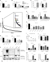
CRISPR-Cas9 screen identifies Cpb1 to be required for caspase-11 expression, activation, and cell death in macrophages. (A) BMDMs were treated with intracellular LPS via CTB for 16 h, and then cytotoxicity was measured. (B) RAW264.7 transformed murine macrophage cell line (RAW) was treated with CTB/LPS or transfected LPS for 16 h and cytotoxicity was measured. (C) Expression of humanized Cas9 (hCas9) was determined in WT RAW cells that were either untreated or transduced with a lentiviral construct that stably expresses hCas (WT-hCas9), or the same cells were treated with LPS/CTB for 16 h and cytotoxicity was measured. (D) A macrophage genetic screen identified KOs that confer resistance to intracellular LPS stimulation via 16 h treatment with CTB/LPS. Each plot represents a gene (x axis). Genes are ranked on the y axis according to the significance (-LogPvalue) of enrichment, which was calculated by an accumulated hypergeometric distribution function (König et al., 2007), in the CTB/LPS-treated library compared with the nonselected control population. The top 100 hits are highlighted in the extrapolated box. Blue genes, CTB receptor genes; purple genes, complement genes; red, yellow, and green, other hits followed up on. (E–I) WT and KO cell lines were treated with LPS/CTB or transfected with LPS for 16 h and cytotoxicity was measured, (I) with or without the Cpb-inhibitor (MGTA). (J) WT, Cpb1 KO, and Cpb1 KO cells complement with either Cpb1 (Cpb1 KO + Cpb1) or caspase-11 (Cpb1 KO + Casp11) on a transgene were treated with CTB/LPS for 16 h and cytotoxicity was measured. (K) WT and KO cell lines were treated with either LPS or CTB/LPS for 16 h. Supernatants and cell lysates were collected for each sample and detection of pro-caspase-11 (pro-Casp11), cleaved caspase-11 (p30 Casp11), and β-actin was evaluated via Western blot. Indicated cell lines were treated with LPS for 2 h. Relative expression of (L) caspase-11 and caspase-1 or (M) IL-6 and TNF were determined. All relative expression was calculated via qRT-PCR, compared with GAPDH expression. All cytotoxicity was measured by release of LDH. Statistics analyzed via the unpaired Student’s t test. *, P < 0.05; **, P < 0.01; ***, P < 0.001; ****, P < 0.0001. Data are representative of at least two (D) or three (A–C and E–M) independent experiments with three technical replicates each time.
Figure 2.
Figure 2.
Complement amplifies TLR4-dependent transcripts and caspase-11–dependent cell death in macrophages. WT, C3aR KO, and C3 KO RAW cell lines were treated with LPS for 2 h and relative expression of (A) caspase-11 or (B) il-6 and tnf was calculated via qRT-PCR, compared with GAPDH expression. Indicated cell lines were treated with CTB/LPS for 16 h and (C) cytotoxicity was measured or (D) supernatants and cell lysates were collected for Western blot analysis of indicated proteins. (E) WT (control [WT] or C3aR-antagonist treated [C3aR-Ant]), caspase-11–deficient (Casp11−/−), C3aR-deficient (C3aR−/−), and C3-deficient (C3−/−) primary BM-derived macrophages (BMDMs) were treated with CTB/LPS for 16 h and cytotoxicity was measured. Indicated BMDMs were treated with LPS for 2 h and the relative expression of (F) caspase-11 or (G) il-6 and tnf was calculated via qRT-PCR, compared with GAPDH expression or (H) analyzed for release of IL-6 and TNF via ELISA. All cytotoxicity was measured by release of LDH. Statistics analyzed via the unpaired Student’s t test. *, P < 0.05; **, P < 0.01; ***, P < 0.001. Data are representative of at least three independent experiments (A–H), with three technical replicates each time.
Figure 3.
Figure 3.
Cpb1–C3–C3aR pathway increases p38 MAPK phosphorylation downstream of TLR4 and Ifnar activation in a cell autonomous and non–cell autonomous manner. WT, C3aR−/−, and C3−/− BMDMs were treated with (A) IFN-β or (B) IFN-γ for 2 h and the relative expression of caspase-11 and NOS2 transcripts were calculated via qRT-PCR, compared with GAPDH expression. (C) WT or C3aR−/− BMDMs were treated with LPS, IFN-β, or IFN-γ for 2 h and lysed for analysis of p38-phosphorylation and β-actin was evaluated via Western blot. (D) WT, Cpb1 KO, or C3aR KO RAW macrophages were treated with LPS for 2 h and supernatants were added to naive Cpb1 KO cells for 2 h. The relative expression of caspase-11, il-6, and tnf-α were calculated via qRT-PCR, compared with GAPDH expression. Statistics were analyzed via the unpaired Student’s t test. *, P < 0.05; ***, P < 0.001; ****, P < 0.0001. Data are representative of at least two (D) or three (A–C) independent experiments, with three technical replicates each time.
Figure 4.
Figure 4.
Cpb1–C3–C3aR pathway is required for caspase-11–dependent cell death in response to Gram-negative bacterial pathogens. Indicated RAW macrophage cell lines were infected with (A) Salmonella Typhimurium (SL1344; MOI 100:1) or (B) S. flexneri (M90T; MOI 50:1) for 18 h and cytotoxicity was measured. (C) BMDMs were infected with S. Typhimurium (SL1344 ΔorgA/fliC; MOI, 100:1) for 18 h and cytotoxicity was measured. All cytotoxicity was measured by release of LDH. Statistics analyzed via the unpaired Student’s t test. *, P < 0.05; **, P < 0.01; ***, P < 0.001. Data are representative of at least three independent experiments (A–C), with three technical replicates each time.
Figure 5.
Figure 5.
C3aR contributes to amplification of proinflammatory mediators in the blood and to the severity of endotoxemia in mice. WT or C3aR-deficient (C3aR−/−) BALBc mice were treated with a sublethal dose of LPS i.p. 2 h pt blood samples were collected and analyzed for expression of (A) caspase-11 or (B) IL-6. Fold-change of gene expression was calculated (Ctrl/LPS treated mice). 6 h pt, mice were treated with a lethal dose of LPS. Weight (C) and temperature (D) were monitored every 2 h for 20 h. Six control mice and eight treated mice were used. Next, WT mice were treated with the same regimen as described with or without C3aRi. As described above, 2 h pt (E) caspase-11 or (F) IL-6 expression was analyzed and fold-change of gene expression was calculated (Ctrl/LPS treated mice), and after 6 h mice were treated with a lethal dose of LPS. Weight (G) and temperature (H) were monitored every 2 h for 20 h. (I) WT mice were treated with LPS and with or without C3aRi i.p. 2 h pt, blood was collected via cardiac bleed and analyzed for IL-6 levels via ELISA. (J) WT mice were treated with LPS and with or without C3aRi, and survival was monitored. n = 5 mice/group in each independent experiment (E–J). Statistics analyzed via the unpaired Student’s t test. *, P < 0.05; **, P < 0.01; ***, P < 0.001, ****, P < 0.0001. Data are representative of at least two (I–J) or three experiments (A–H), with three technical replicates each time.
Figure 6.
Figure 6.
C3aR plays a role in enhanced inflammation in primary human macrophages and is expressed with caspase-5 in PMBCs from patients with sepsis. Primary MDMs were (A) untreated or treated with LPS for 1–3 h, or (B) untreated or treated with C3aRi, with and without LPS stimulation, for 3 h, and protein levels of pro-caspase-4, pro-caspase-5, caspase-5 p20, and β-actin were evaluated via Western blot. (C) MDMs were untreated or treated with C3aRi, with and without LPS, IFN-β, or IFN-γ for 3 h, and levels of phosphorylated p-38 or β-actin was evaluated via Western blot. (D) IL-6 and TNF cytokine release from MDMs was assessed via ELISA, 3 h after stimulation with LPS with or without C3aRi. Forest plots for random effects model estimates of effect size of the induction of (E) caspase-5 and C3aR1 genes in healthy controls versus patients with acute infections or sepsis in eight cohorts. Statistics analyzed via the unpaired Student’s t test. *, P < 0.05; **, P < 0.01; ***, P < 0.001. Data are representative of at least three experiments (A–D), with three technical replicates each time.
Figure 7.
Figure 7.
Model of Cpb1–C3–C3aR pathway amplifying p38 MAPK signaling downstream of TLR4 and Ifnar activation in macrophages. TLR4 and Ifnar activation through their respective ligands induce expression of proinflammatory genes via various mechanisms, including the induction of p38 MAPK (Lee et al., 1994; Sancéau et al., 2000). C3 expression is induced downstream these pathways (Riches et al., 1988; Maranto et al., 2011), and is quickly released from macrophages (Goodrum, 1987), where it is cleaved into C3a and C3b. Anaphylotoxin C3a can be cleaved extracellularly by Cpb1 into C3a-desArg (Leung et al., 2008; Chatterjee et al., 2009), where it then acts as a ligand for C3aR. We find, the activation of C3aR then amplifies MAPK activity through enhancing p38 MAPK phosphorylation downstream of TLR4 and Ifnar, but not Ifngr, activation. Due to the extracellular nature of this signaling pathway, the Cpb1–C3–C3aR pathway acts as a cell-autonomous and a non–cell autonomous amplification pathway enhancing expression of proinflammatory genes of self- and neighboring macrophages.

References

    1. Aachoui Y., Leaf I.A., Hagar J.A., Fontana M.F., Campos C.G., Zak D.E., Tan M.H., Cotter P.A., Vance R.E., Aderem A., and Miao E.A.. 2013. Caspase-11 protects against bacteria that escape the vacuole. Science. 339:975–978. 10.1126/science.1230751 - DOI - PMC - PubMed
    1. Ames R.S., Lee D., Foley J.J., Jurewicz A.J., Tornetta M.A., Bautsch W., Settmacher B., Klos A., Erhard K.F., Cousins R.D., et al. . 2001. Identification of a selective nonpeptide antagonist of the anaphylatoxin C3a receptor that demonstrates antiinflammatory activity in animal models. J. Immunol. 166:6341–6348. 10.4049/jimmunol.166.10.6341 - DOI - PubMed
    1. Angus D.C., and van der Poll T.. 2013. Severe sepsis and septic shock. N. Engl. J. Med. 369:2063 10.1056/NEJMra1208623 - DOI - PubMed
    1. Angus D.C., Linde-Zwirble W.T., Lidicker J., Clermont G., Carcillo J., and Pinsky M.R.. 2001. Epidemiology of severe sepsis in the United States: analysis of incidence, outcome, and associated costs of care. Crit. Care Med. 29:1303–1310. 10.1097/00003246-200107000-00002 - DOI - PubMed
    1. Ardura M.I., Banchereau R., Mejias A., Di Pucchio T., Glaser C., Allantaz F., Pascual V., Banchereau J., Chaussabel D., and Ramilo O.. 2009. Enhanced monocyte response and decreased central memory T cells in children with invasive Staphylococcus aureus infections. PLoS One. 4:e5446 10.1371/journal.pone.0005446 - DOI - PMC - PubMed

MeSH terms