. 2016 Nov 15;143(22):4193-4202.
doi: 10.1242/dev.141192. Epub 2016 Oct 3.

Nuclei migrate through constricted spaces using microtubule motors and actin networks in C. elegans hypodermal cells

Affiliations

Nuclei migrate through constricted spaces using microtubule motors and actin networks in C. elegans hypodermal cells

Courtney R Bone et al. Development. .

Abstract

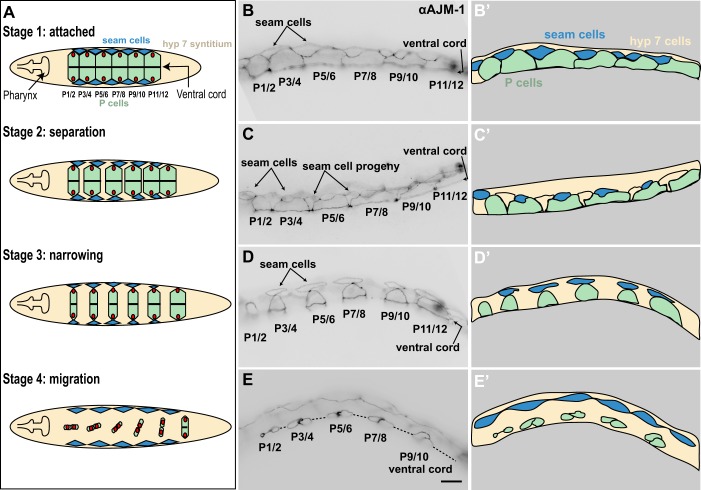
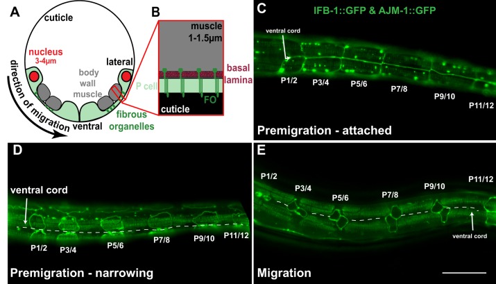
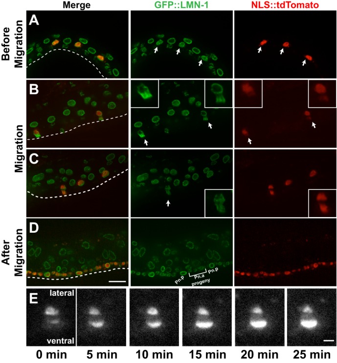
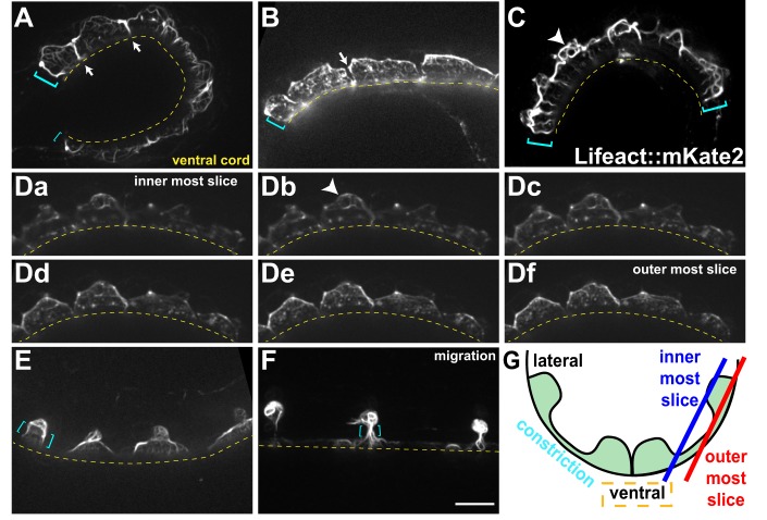
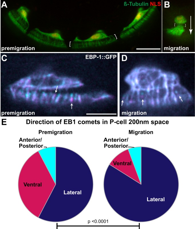
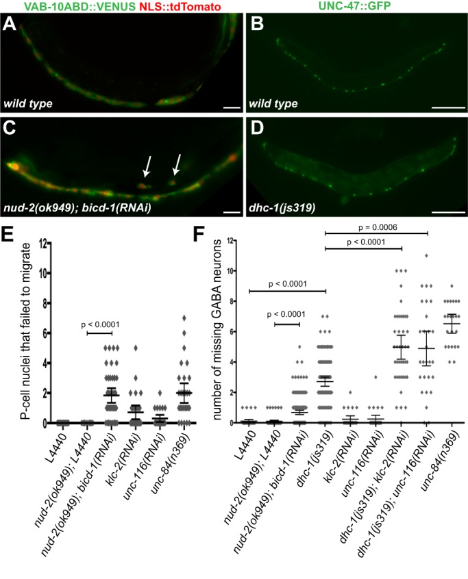
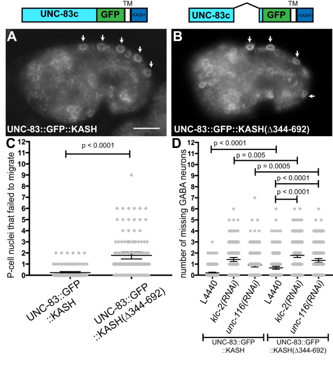
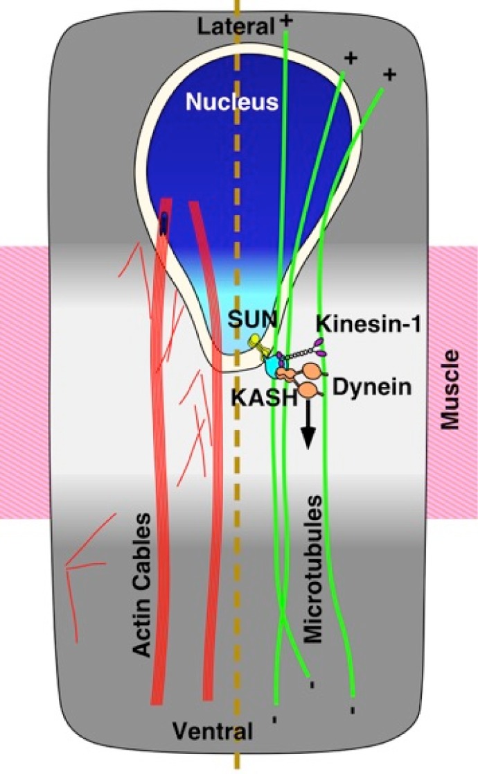
Cellular migrations through constricted spaces are a crucial aspect of many developmental and disease processes including hematopoiesis, inflammation and metastasis. A limiting factor in these events is nuclear deformation. Here, we establish an in vivo model in which nuclei can be visualized while moving through constrictions and use it to elucidate mechanisms for nuclear migration. C. elegans hypodermal P-cell larval nuclei traverse a narrow space that is about 5% their width. This constriction is blocked by fibrous organelles, structures that pass through P cells to connect the muscles to cuticle. Fibrous organelles are removed just prior to nuclear migration, when nuclei and lamins undergo extreme morphological changes to squeeze through the space. Both actin and microtubule networks are organized to mediate nuclear migration. The LINC complex, consisting of the SUN protein UNC-84 and the KASH protein UNC-83, recruits dynein and kinesin-1 to the nuclear surface. Both motors function in P-cell nuclear migration, but dynein, functioning through UNC-83, plays a more central role as nuclei migrate towards minus ends of polarized microtubule networks. Thus, the nucleoskeleton and cytoskeleton are coordinated to move nuclei through constricted spaces.

Keywords: C. elegans; Dynein; KASH; Nuclear migration; SUN.


Conflict of interest statement

The authors declare no competing or financial interests.

Figures

Fig. 1.
Fig. 1.
Stages of P-cell morphology throughout early L1 development. Four stages of P-cell morphological changes are shown. (A) Three cell types are depicted in ventral view: P cells (green), the hyp7 syncytium (tan) and seam cells (blue). Stage 1, attached: 12 P cells organized in six pairs with their nuclei (red) on the lateral side. P cells are attached to their anterior/posterior neighbors to cover the ventral surface completely. Stage 2, separation: P cells separate from their neighbors and hyp7 cells fill the openings. Stage 3, narrowing: P cell pairs narrow and hyp7 cells expand to fill the resulting spaces. Stage 4, migration: Nuclei migrate from lateral to ventral. Anterior pairs of P cells migrate first. By the end of migration the P-cell cytoplasm retracts to the ventral cord. (B-E) Lateral views of the four stages of fixed larvae stained with an anti-AJM-1 antibody to mark adherens junctions. (B′-E′) The same images pseudocolored to identify cell types. Scale bar: 10 µm.
Fig. 2.
Fig. 2.
Fibrous organelles are disassembled in P cells at the time of nuclear migration. (A) A cross-section depicting the shape of P cells. The P cell (green) is constricted between the cuticle (black) and body wall muscles (gray), and the nuclei (red) are on the lateral sides. (B) The cuticle is connected to the body wall muscle through P cells and the basal lamina (maroon) by fibrous organelles (FO, green). (C-E) Animals were visualized expressing both AJM-1::GFP and IFB-1::GFP to visualize fibrous organelles. Large aggregates, particularly in C, are an artifact of AJM-1::GFP overexpression. (C) Stage 1, attached. Fibrous organelles are seen ‘end on’, but line up close to one another and appear in lines. (D) Stage 3, narrowing. Fibrous organelles in P cells (outlined by AJM-1::GFP) and hyp7 between P cells. (E) Stage 4, nuclear migration. Fibrous organelles are absent in P cells during migration (P3/4) and are in the process of disassembling just prior to migration (P7/8). Fibrous organelles are reorganized in the hyp7 syncytium following P-cell cytoplasm retraction to the ventral cord (visible at P1/2). Dashed line represents the ventral cord. Scale bar: 20 µm.
Fig. 3.
Fig. 3.
P-cell nuclei squeeze through constricted spaces. Images of larvae with NLS::tdTomato in P-cell lineages (red, or white in E) and endogenously expressed GFP::LMN-1 (green). All images are lateral views of L1 larvae with ventral (dashed line) down and anterior left. (A) A larva prior to nuclear migration with GFP::LMN-1 surrounding all visible nuclei. Arrows mark P-cell nuclei. (B,C) Larvae at the time of P-cell nuclear migration. P-cell nuclei that are undergoing migration are marked by arrows. Insets show enlarged images of P-cell nuclei during migration. (D) A larva after P-cell migration. (E) A single migrating P-cell nucleus shown over a 25 min period. Images are 5 min apart. Scale bars: 10 µm (A-D); 5 µm (E).
Fig. 4.
Fig. 4.
Actin networks in larval P cells. (A-F) Lifeact::mKate2 expressed in P cells is shown. Lateral views with ventral (dashed line) down and anterior left. Pre-migration stage larval P cells have either thick actin cables (A,C) or thin actin filaments (B,D). Actin is enriched at the cell periphery and cell-cell boundaries (arrows in A,B). Actin rings are seen in some larvae (arrowheads C,Db). (Da-f) A single premigration larva is shown in six images from a z-stack (200 nm steps) starting with an interior slice showing the ventral cord followed by images moving up and out to the lateral side. (E) Actin cables and peripheral enrichment in narrowing cells. (F) Actin cables visualized through the constriction (cyan brackets) in migration stage P cells. (G) Schematic depicting a P cell (green) cross-section labeled with the ventral side (yellow dashed line in images), lateral side, and the constriction (cyan brackets in images). Inner and outer boundaries of the z-stack shown in D are marked.
Fig. 5.
Fig. 5.
Microtubules are polarized at the time of P-cell nuclear migration. (A,B) Larvae expressing β-tubulin::GFP and NLS::tdTomato in P cells. Lateral views (ventral down, anterior left) are shown and brackets mark the constrictions. Scale bar: 10 µm. (A) Four premigration cells are shown. (B) Single cell at the time of nuclear migration. Migration occurs from top to bottom, see arrow. (C,D) P cells expressing EBP-1::GFP were imaged every second for up to 3 minutes. Two frames, 5 s apart are shown with the first image pseudocolored magenta and the second cyan to show the direction of growth. Example EB1 comets in the constricted space are marked with arrows showing the direction of growth. Scale bar: 5 µm. (C) A premigration P cell at separation stage. (D) A P cell about to migrate. (E) Graphs showing the percentage of EB1 comets in the constricted space growing towards the lateral side (dark blue), the ventral side (magenta) or towards the anterior and posterior (perpendicular to axis of migration, cyan) (491 premigration comets from ten cells and 405 comets from 12 migration stage cells were analyzed). The two populations of EB1 comets are significantly different (P<0.0001 by χ2 contingency test).
Fig. 6.
Fig. 6.
Dynein and kinesin function to move P-cell nuclei. (A,C) Wild-type (A) and nud-2(ok949);bicd-1(RNAi) (C) late L1 larvae expressing P cell-specific (hlh-3 promoter-driven) VAB-10ActinBindingDomain::VENUS and NLS::tdTomato. Arrows mark nuclei on the lateral side of nud-2(ok949); bicd-1(RNAi) larva that failed to migrate. Scale bars: 10 µm. (B,D) Wild-type (B) and dhc-1(js319) (D) young adults expressing UNC-47::GFP. Lateral views, ventral down and anterior left. Scale bars: 100 µm. (E,F) Graphs of animals assayed for P-cell nuclear migration failure. Each diamond represents the number of P-cell nuclei that failed to migrate in a single animal. The bars mark mean and 95% CI. (E) Larvae assayed after the time of nuclear migration for the presence of failed P-cell nuclei on the lateral side. (F) Animals assayed as L4 larvae and young adults for the number of GABA neurons missing.
Fig. 7.
Fig. 7.
UNC-83 is required to recruit dynein for P-cell nuclear migration. (A,B) Images of comma-stage embryos show normal UNC-83 localization. Arrows mark UNC-83-positive hyp7 nuclei. (A) An animal expressing UNC-83::GFP::KASH. (B) UNC-83(Δ344-692)::GFP::KASH mutant embryo. Ventral down and anterior left. Scale bar: 10 µm. (C) Graph showing larvae assayed for P-cell nuclear migration defects. (D) Graph of the number of GABA neurons missing in L4 larvae and young adult animals. Each diamond on the graphs represents a single animal; bars mark mean and 95% CI.
Fig. 8.
Fig. 8.
Model of P-cell nuclear migration. The P-cell nucleus (blue) migrates from the lateral (top) to ventral (bottom) through a constricted space (light gray) between the muscle (pink) and the cuticle (not shown, but above the plane of the image). P-cell nuclear migration relies on a microtubule pathway consisting of polarized microtubules (green) and SUN (yellow), KASH (cyan), kinesin (purple) and dynein (tan) at the nuclear envelope. Actin cables (red) extend throughout the P cell.

Similar articles

Cited by

References

    1. Altun Z. F. and Hall D. H. (2009a). Epithelial system, hypodermis. WormAtlas, doi:10.3908/wormatlas.1.13 10.3908/wormatlas.1.13 - DOI
    1. Altun Z. F. and Hall D. H. (2009b). Muscle system, somatic muscle. WormAtlas, doi:10.3908/wormatlas.1.7 10.3908/wormatlas.1.7 - DOI
    1. Arribere J. A., Bell R. T., Fu B. X. H., Artiles K. L., Hartman P. S. and Fire A. Z. (2014). Efficient marker-free recovery of custom genetic modifications with CRISPR/Cas9 in Caenorhabditis elegans. Genetics 198, 837-846. 10.1534/genetics.114.169730 - DOI - PMC - PubMed
    1. Bolhy S., Bouhlel I., Dultz E., Nayak T., Zuccolo M., Gatti X., Vallee R., Ellenberg J. and Doye V. (2011). A Nup133-dependent NPC-anchored network tethers centrosomes to the nuclear envelope in prophase. J. Cell Biol. 192, 855-871. 10.1083/jcb.201007118 - DOI - PMC - PubMed
    1. Bone C. R., Tapley E. C., Gorjanacz M. and Starr D. A. (2014). The C. elegans SUN protein UNC-84 interacts with lamin to transfer forces from the cytoplasm to the nucleoskeleton during nuclear migration. Mol. Biol. Cell 25, 2853-2865. 10.1091/mbc.E14-05-0971 - DOI - PMC - PubMed

Publication types