. 2016 Oct 10;215(1):95-106.
doi: 10.1083/jcb.201603062. Epub 2016 Oct 3.

ASB7 regulates spindle dynamics and genome integrity by targeting DDA3 for proteasomal degradation

Affiliations

ASB7 regulates spindle dynamics and genome integrity by targeting DDA3 for proteasomal degradation

Keiji Uematsu et al. J Cell Biol. .

Abstract

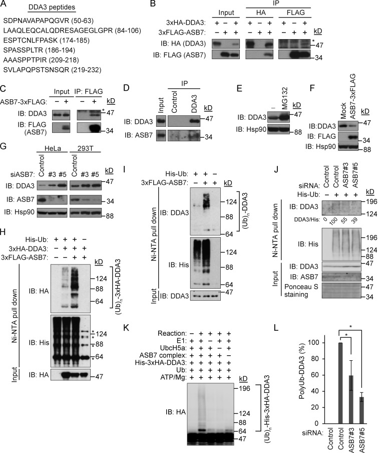
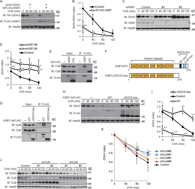
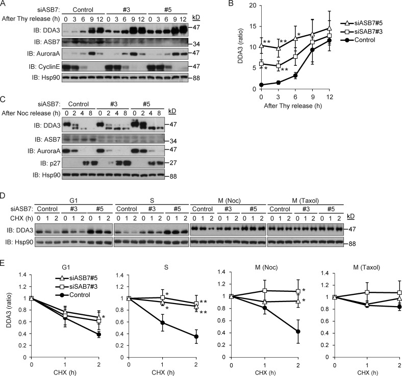
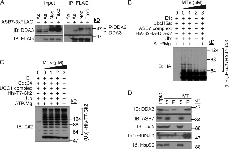
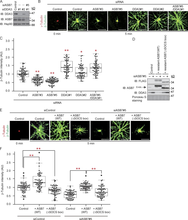
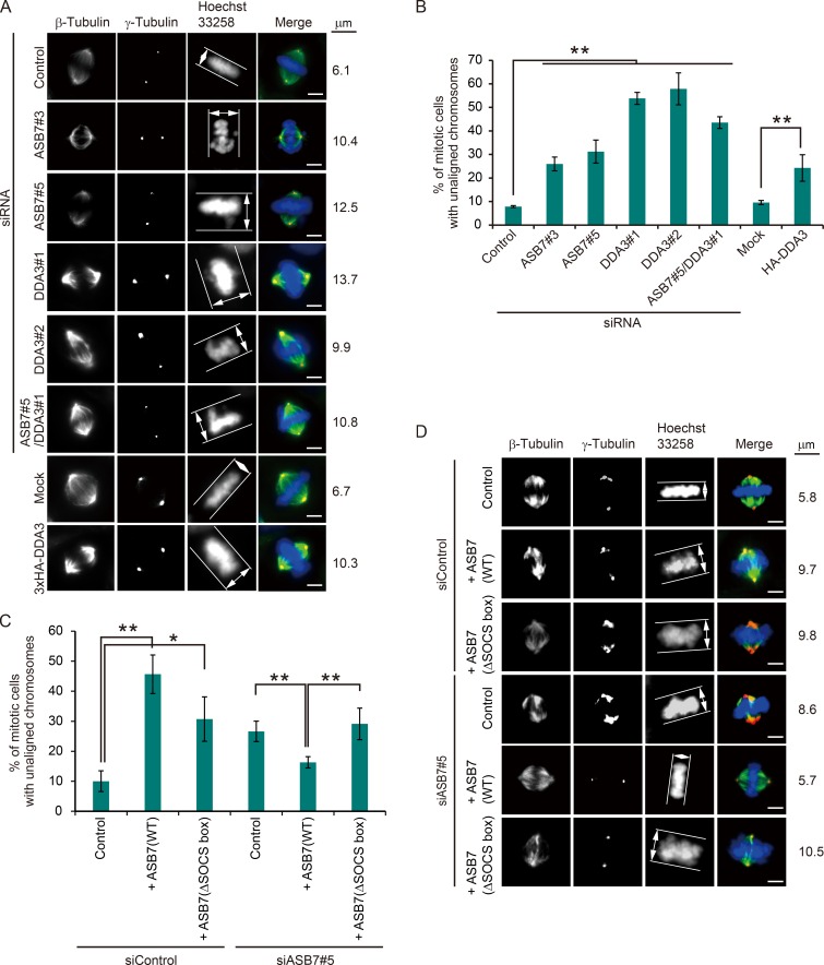
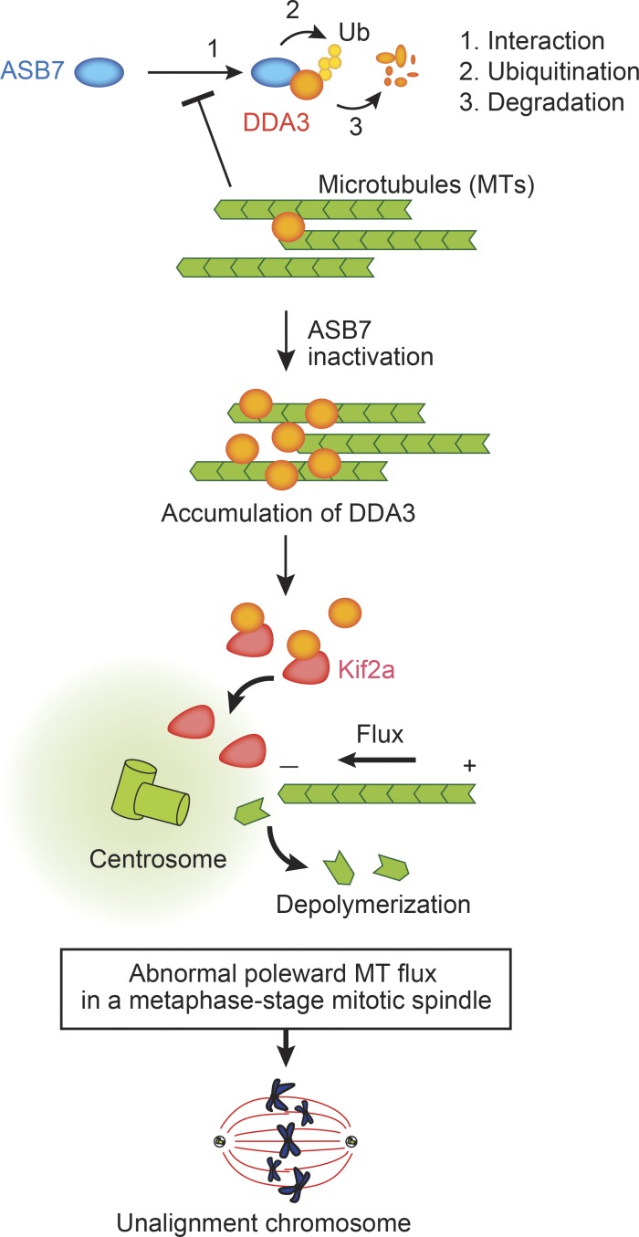
Proper dynamic regulation of the spindle is essential for successful cell division. However, the molecular mechanisms that regulate spindle dynamics in mitosis are not fully understood. In this study, we show that Cullin 5-interacting suppressor of cytokine signaling box protein ASB7 ubiquitinates DDA3, a regulator of spindle dynamics, thereby targeting it for proteasomal degradation. The presence of microtubules (MTs) prevented the ASB7-DDA3 interaction, thus stabilizing DDA3. Knockdown of ASB7 decreased MT polymerization and increased the proportion of cells with unaligned chromosomes, and this phenotype was rescued by deletion of DDA3. Collectively, these data indicate that ASB7 plays a crucial role in regulating spindle dynamics and genome integrity by controlling the expression of DDA3.


Figures

Figure 1.
Figure 1.
ASB7 interacts with DDA3 and promotes ubiquitination of DDA3. (A) Identification of DDA3 as an ASB7-interacting protein. 3×FLAG-ASB7 expressed in HEK293T cells was purified using anti-FLAG antibody and resolved by SDS-PAGE. ASB7-interacting proteins were analyzed by mass spectrometry. Identified peptides of DDA3 are shown. (B) Interaction between 3×HA-DDA3 and 3×FLAG-ASB7. HEK293T cells expressing 3×HA-DDA3 and 3×FLAG-ASB7 (as indicated) were cultured in the presence of the proteasome inhibitor MG132 (10 µM for 6 h), immunoprecipitated (IP) with anti-HA or anti-FLAG antibody, and immunoblotted (IB) with anti-HA or anti-FLAG antibody. (C) Interaction between endogenous DDA3 and 3×FLAG-ASB7. HeLa cells stably expressing 3×FLAG-ASB7 were cultured in the presence of MG132 (10 µM for 6 h), immunoprecipitated with anti-FLAG antibody, and immunoblotted with anti-DDA3 or anti-FLAG antibody. (D) Interaction between endogenous DDA3 and ASB7. HeLa cells were cultured in the presence of MG132 (10 µM for 6 h), immunoprecipitated with anti-DDA3 antibody, and immunoblotted with anti-DDA3 or anti-ASB7 antibody. (E) Accumulation of DDA3 upon exposure to MG132. HeLa cells were cultured in the presence or absence of 10 µM MG132 for 6 h and then Western blotted with antibodies against DDA3. (F) Down-regulation of DDA3 by overexpression of ASB7. HeLa cells stably expressing 3×FLAG-ASB7 were lysed and immunoblotted with anti-DDA3, anti-FLAG, or anti-Hsp90 antibody. (G) Accumulation of endogenous DDA3 by knockdown of ASB7. Two independent siRNAs (#3 and #5) targeting ASB7 were transfected into HeLa or HEK293T cells, which were cultured for 2 d and then Western blotted with anti-DDA3, anti-FLAG, or anti-Hsp90 antibody. (E–G) Hsp90 is shown as a loading control. (H) ASB7-dependent polyubiquitination of exogenous DDA3 in vivo. HEK293T cells were transfected with plasmids encoding 3×FLAG-ASB7, 3×HA-DDA3, and/or His6-tagged ubiquitin. MG132 (10 µM for 6 h) was used to detect polyubiquitination. Cell lysates were subjected to nickel–nitrilotriacetic acid (Ni-NTA) pulldown to purify proteins modified by His6-ubiquitin, followed by IB analysis with anti-HA, FLAG, or His6 antibody. Asterisks denote nonspecific bands. (I) ASB7-dependent polyubiquitination of endogenous DDA3 in vivo. HeLa cells stably expressing 3×FLAG-ASB7 were examined as in H. (J) Reduced polyubiquitination of endogenous DDA3 by knockdown of ASB7. siRNAs (#3 and #5) targeting ASB7 were transfected into HEK293T cells and examined as in I. (K) ASB7-dependent polyubiquitination of DDA3 in vitro. Recombinant ubiquitin-activating enzyme (E1), ubiquitin-conjugating enzyme (UbcH5a), ASB7 complex (ASB7, Cul5, Elongin B, Elongin C, and Rbx2), His6-ubiquitin, and immunopurified 3×HA-DDA3 were mixed in vitro in various combinations in the presence of ATP, incubated at 28°C for 1 h, and subjected to Western blotting with anti-HA antibody. (L) Quantification of polyubiquitinated DDA3. The signals of polyubiquitinated DDA3 shown in J were quantified. Control sample with or without His6-ubiquitin expression was set as 100 and 0, respectively. *, P < 0.02. Data represent the mean ± SD of three independent experiments.
Figure 2.
Figure 2.
Destabilization of DDA3 by ASB7. (A) HEK293T cells expressing 3×HA-DDA3 with or without 3×FLAG-ASB7 were exposed to 50 µg/ml CHX for 40, 80, or 120 min. The lysates were subjected to Western blotting with antibodies against HA, FLAG, or Hsp90. (B) The intensities of HA-DDA3 bands in A were normalized to those of the corresponding Hsp90 bands and plotted as a ratio of the normalized value without ASB7 expression at 0 h. (C) Stabilization of endogenous DDA3 by knockdown of ASB7. Two independent siRNAs (#3 and #5) targeting ASB7 were transfected into HeLa cells and cultured for 2 d, followed by CHX treatment as in A. The lysates were subjected to Western blotting with antibodies against DDA3, ASB7, or Hsp90. (D) The intensities of DDA3 bands in C were normalized to those of the corresponding Hsp90 bands and plotted as a ratio of the normalized value of control cells at 0 h. (E) ASB7 interacts with Cul5 but not Cul2. HEK293T cells expressing 3×FLAG-ASB7 or 3×FLAG-pVHL (Cul2-type ubiquitin ligase as a control) were cultured in the presence of MG132 (10 µM for 6 h), immunoprecipitated (IP) with anti-FLAG antibody, and immunoblotted (IB) with anti-Cul2, anti-Cul5, or anti-FLAG antibody. (F) Schematic representation of ASB7 mutants used in this study. (G) Interaction between the SOCS box of ASB7 and Cul5. HeLa cells stably expressing 3×FLAG-ASB7(WT or ΔSOCS box) were lysed, immunoprecipitated with anti-FLAG antibody, and immunoblotted with anti-Cul2, anti-Cul5, or anti-FLAG antibody. (E and G) Asterisk denotes a nonspecific band. (H) Inactivation of ASB7 by deletion of the SOCS box. HeLa cells stably expressing 3×FLAG-ASB7(WT or ΔSOCS box) were exposed to CHX as in A. The lysates were subjected to Western blotting with antibodies against DDA3, FLAG, or Hsp90. (I) The intensities of DDA3 bands in H were normalized to those of the corresponding Hsp90 bands and plotted as a ratio of the normalized value of control cells at 0 h. (J) Endogenous DDA3 was stabilized by knockdown of Cul5, but not Cul2. Two independent siRNAs (#1 and #2) targeting Cul2 or Cul5 were transfected into HeLa cells and cultured for 2 d, followed by CHX treatment as in A. The lysates were subjected to Western blotting with antibodies against DDA3, Cul2, Cul5, or Hsp90. (A, C, H, and J)_Hsp90 is shown as a loading control. (K) The intensities of DDA3 bands in J were normalized to those of the corresponding Hsp90 bands and plotted as a ratio of the normalized value of control cells at 0 h. For all graphs, *, P < 0.05; **, P < 0.01. Data represent the mean ± SD of three independent experiments (B, D, and I) and four independent experiments (K).
Figure 3.
Figure 3.
Cell cycle–dependent regulation of DDA3 by ASB7. (A) Stabilization of DDA3 in S phase by knockdown of ASB7. Two independent siRNAs (#3 and #5) targeting ASB7 were transfected into HeLa cells and synchronized in S phase. The cells were released and harvested at the indicated times. The lysates were subjected to Western blotting with antibodies against DDA3, ASB7, Aurora A, cyclin E, or Hsp90. (B) The intensities of DDA3 bands in A were normalized to those of the corresponding Hsp90 bands and plotted as a ratio of the normalized value of control cells at 0 h. (C) Knockdown of ASB7 had no effect on the stability of DDA3 in M phase. Two independent siRNAs (#3 and #5) targeting ASB7 were transfected into HeLa cells and synchronized in M phase. The cells were released and harvested at the indicated times. The lysates were subjected to Western blotting with antibodies against DDA3, ASB7, Aurora A, p27, or Hsp90. (D) ASB7-dependent destabilization of DDA3 in S phase. Two independent siRNAs (#3 and #5) targeting ASB7 were transfected into HeLa cells and synchronized in the G1, S, or M phase. Nocodazole or taxol was used to synchronize cells in M phase. The cells were then exposed to 50 µg/ml CHX for 1 or 2 h. The lysates were subjected to Western blotting with antibodies against DDA3 or Hsp90. (A, C, and D) Hsp90 is shown as a loading control. (E) The intensities of DDA3 bands in D were normalized to those of the corresponding Hsp90 bands and plotted as a ratio of the normalized value of control cells at 0 h. For all graphs, *, P < 0.05; **, P < 0.01. Data represent the mean ± SD of three independent experiments.
Figure 4.
Figure 4.
Prevention of polyubiquitination of DDA3 by MTs. (A) Interaction between ASB7 and nonphosphorylated or phosphorylated DDA3. HeLa cells stably expressing 3×FLAG-ASB7 or control cells were treated with 0.1 µg/ml nocodazole or 1 µM taxol for 16 h. Cells were further incubated in the presence of 10 µM MG132 for 4 h, and suspended cells in the culture medium were lysed, immunoprecipitated (IP) with anti-FLAG antibody, and immunoblotted (IB) with anti-DDA3 or anti-FLAG antibody. As, asynchronized. (B) Prevention of polyubiquitination of DDA3 by MTs in a dose-dependent manner. Different amounts of MTs were added to the in vitro polyubiquitination reactions. (C) MTs had no effect on polyubiquitination of Cit2. Different amounts of MTs were added to the in vitro polyubiquitination reactions. (D) ASB7 does not interact with MTs. Whole-cell lysates (input) of HeLa cells were prepared and incubated with or without taxol-stabilized MTs. Samples were centrifuged, and supernatant (S) and pellet (P) fractions were analyzed by immunoblotting with antibodies against DDA3, ASB7, Cul5, α-tubulin, or Hsp90. Hsp90 does not bind to MTs and was used as a negative control.
Figure 5.
Figure 5.
Regulation of MT polymerization by ASB7 and DDA3. (A) Knockdown of ASB7 and/or DDA3. Two independent siRNAs (#1 and #2) targeting DDA3 or siRNA targeting ASB7 were transfected into HeLa cells. The lysates were subjected to Western blotting with antibodies against DDA3, ASB7, or Hsp90. Hsp90 is shown as a loading control. Asterisk denotes a nonspecific band. (B) MT regrowth assay using ASB7 and/or DDA3 knockdown HeLa cells. After incubation for 5 min to allow MT regrowth, cells were fixed and immunostained with anti–β-tubulin or anti–γ-tubulin. (C) Box plots of β-tubulin intensities after MT regrowth. About 20 cells from five randomly selected loci were examined in each experiment. The experiment was repeated three times independently, and a total of 60 cells were examined. The mean of normalized fluorescence intensity, expressed in arbitrary units (AU) of fluorescence, was calculated. The box plot shows the normalized expression values: bottom line outside the box, 10th percentile; bottom border of the box, 25th percentile; middle line within the box, median; upper border of the box, 75th percentile; top line outside the box, 90th percentile. Each dot indicates the intensity of an individual cell. (D) Complementation of ASB7 in ASB7 knockdown cells prevents DDA3 accumulation. siRNA targeting ASB7 or control was transfected into HeLa cells with stably expressing siRNA-resistant ASB7-3×FLAG (WT or ΔSOCS box). The lysates were subjected to Western blotting with antibodies against FLAG, ASB7, or DDA3. Ponceau S staining is shown as a loading control. Endogenous ASB7 is indicated by an arrow. (E) MT regrowth assay using ASB7 knockdown or ASB7-complemented HeLa cells performed as in B. Bars, 5 µm. (F) Box plots of β-tubulin intensities after MT regrowth calculated as in C. For all graphs, *, P < 0.05; **, P < 0.01.
Figure 6.
Figure 6.
Regulation of chromosome alignment by ASB7 and DDA3. (A) Maximum projections from deconvolved z stacks of representative ASB7, DDA3, and/or Kif2a knockdown HeLa cells or HeLa cells stably overexpressing 3×HA-DDA3, as well as control cells, immunostained for β- or γ-tubulin. DNA was stained with Hoechst 33258. (B) Proportion of mitotic cells with unaligned chromosomes. More than 150 mitotic HeLa cells were counted, and percentages of cells with unaligned chromosomes are shown. (C) Proportion of mitotic cells with unaligned chromosomes. More than 150 mitotic HeLa cells were counted, and percentages of cells with unaligned chromosomes are shown. *, P < 0.05; **, P < 0.01. Data represent means ± SD. (D) Maximum projections from deconvolved z stacks of representative ASB7 knockdown or ASB7-complemented HeLa cells, immunostained for β- or γ-tubulin. DNA was stained with Hoechst 33258. Bars, 5 µm.
Figure 7.
Figure 7.
Model of regulation of cell division by ASB7. ASB7 targets DDA3 for polyubiquitination and proteasomal degradation. MTs prevent interaction between ASB7 and DDA3. Inactivation or loss of ASB7 results in accumulation of DDA3, which influences MT flux in collaboration with Kif2a. The abnormal poleward MT flux impedes normal chromosome alignment in metaphase.

Similar articles

Cited by

References

    1. Cai S., O’Connell C.B., Khodjakov A., and Walczak C.E.. 2009. Chromosome congression in the absence of kinetochore fibres. Nat. Cell Biol. 11:832–838. 10.1038/ncb1890 - DOI - PMC - PubMed
    1. Eagleson G., Pfister K., Knowlton A.L., Skoglund P., Keller R., and Stukenberg P.T.. 2015. Kif2a depletion generates chromosome segregation and pole coalescence defects in animal caps and inhibits gastrulation of the Xenopus embryo. Mol. Biol. Cell. 26:924–937. 10.1091/mbc.E13-12-0721 - DOI - PMC - PubMed
    1. Ems-McClung S.C., and Walczak C.E.. 2010. Kinesin-13s in mitosis: Key players in the spatial and temporal organization of spindle microtubules. Semin. Cell Dev. Biol. 21:276–282. 10.1016/j.semcdb.2010.01.016 - DOI - PMC - PubMed
    1. Gaedcke J., Grade M., Jung K., Camps J., Jo P., Emons G., Gehoff A., Sax U., Schirmer M., Becker H., et al. . 2010. Mutated KRAS results in overexpression of DUSP4, a MAP-kinase phosphatase, and SMYD3, a histone methyltransferase, in rectal carcinomas. Genes Chromosomes Cancer. 49:1024–1034. 10.1002/gcc.20811 - DOI - PMC - PubMed
    1. Ganem N.J., and Compton D.A.. 2004. The KinI kinesin Kif2a is required for bipolar spindle assembly through a functional relationship with MCAK. J. Cell Biol. 166:473–478. 10.1083/jcb.200404012 - DOI - PMC - PubMed

Associated data