Review
. 2016 Oct 3;8(10):a006072.
doi: 10.1101/cshperspect.a006072.

Cell Signaling and Stress Responses

Affiliations
Review

Cell Signaling and Stress Responses

Gökhan S Hotamisligil et al. Cold Spring Harb Perspect Biol. .

Abstract

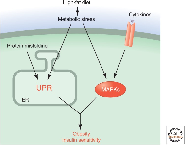
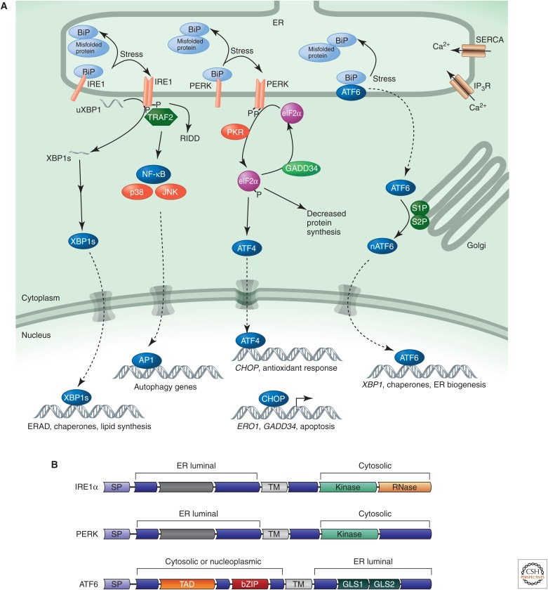
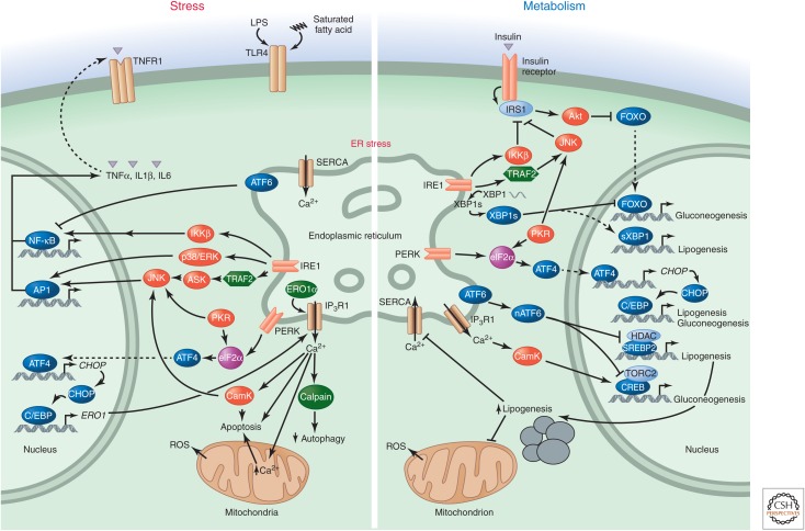
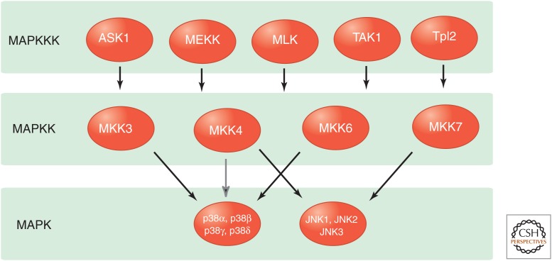
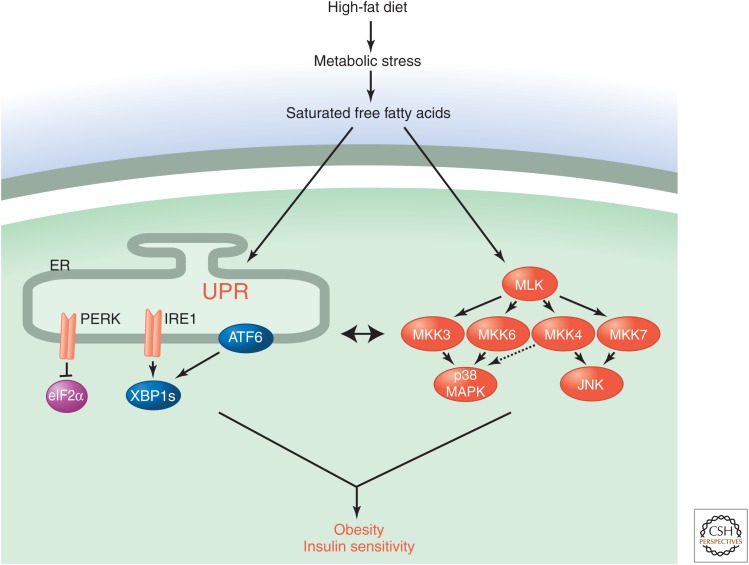
Stress-signaling pathways are evolutionarily conserved and play an important role in the maintenance of homeostasis. These pathways are also critical for adaptation to new cellular environments. The endoplasmic reticulum (ER) unfolded protein response (UPR) is activated by biosynthetic stress and leads to a compensatory increase in ER function. The JNK and p38 MAPK signaling pathways control adaptive responses to intracellular and extracellular stresses, including environmental changes such as UV light, heat, and hyperosmotic conditions, and exposure to inflammatory cytokines. Metabolic stress caused by a high-fat diet represents an example of a stimulus that coordinately activates both the UPR and JNK/p38 signaling pathways. Chronic activation of these stress-response pathways ultimately causes metabolic changes associated with obesity and altered insulin sensitivity. Stress-signaling pathways, therefore, represent potential targets for therapeutic intervention in the metabolic stress response and other disease processes.


Figures

Figure 1.
Figure 1.
Stress-signaling pathways activated in response to metabolic stress. Feeding mice a high-fat diet causes metabolic stress that leads to the UPR and activation of stress-activated MAP kinases. These signaling pathways result in an adaptive response associated with obesity and altered insulin sensitivity.
Figure 2.
Figure 2.
The canonical UPR. (A) In the canonical model of the UPR, unfolded or misfolded proteins activate the three major sensing molecules (IRE1, PERK, and ATF6) at the ER membrane by recruiting the ER chaperone BiP away from the lumenal domains of these proteins. IRE1 is a kinase and ribonuclease that on autophosphorylation activates splicing and produces the active transcription factor XBP1, which induces the expression of ER chaperones, degradation components, and lipid synthesis enzymes. PERK is a kinase that is also activated through dimerization and autophosphorylation and phosphorylates eIF2α to attenuate general protein synthesis. ATF6 is a transcription factor that once released from the ER will move to the Golgi. After processing at this site, it translocates to the nucleus to activate the transcription of chaperone genes. Together, these pathways reduce entry of proteins into the ER, facilitate disposal of the misfolding proteins, and produce the components for the ER to adapt its folding capacity to reach equilibrium. When these pathways fail to reach homeostasis, they can also trigger death. Under severe stress conditions, the synthesis of ATF4 is enhanced in an eIF2α-phosphorylation-independent manner that promotes apoptosis. (B) Domain structure of the ER stress sensors IRE1, PERK, and ATF6. SP, signal peptide; TM, transmembrane domain; TAD, transcriptional activation domain; bZIP, basic leucine zipper; GLS1 and GLS2, Golgi localization sequences 1 and 2. Dark gray bars represent regions of limited sequence similarity between IRE1 and PERK.
Figure 3.
Figure 3.
The UPR in stress signaling, inflammation, and metabolism. The UPR contributes to both inflammatory/stress signaling and metabolic regulation, as exemplified in chronic metabolic diseases. It activates several stress-related kinases including ERK, p38, JNK, and IKK. The resulting signals can impair signaling by insulin or other endocrine hormones and disrupt metabolism. The UPR can also modulate glucose and lipid metabolism directly. Nuclear ATF6 inhibits gluconeogenesis and lipogenesis by directly binding to TORC2 and SREBP2 proteins, respectively. This mechanism is defective in metabolic disease. The spliced form of XBP1 (XBP1s) can directly or indirectly (through SREBP1) activate the lipogenesis program while inhibiting gluconeogenesis. eIF2α phosphorylation leads to the synthesis of CHOP and the activation of lipogenesis programs via CEBPα/β. (Note that the opposing effects of the UPR on gluconeogenesis and lipogenesis are context dependent.) Activation of lipogenesis alters the membrane lipid composition of the ER, inhibits SERCA calcium pumps, and propagates ER stress, which in turn disrupts metabolism further. Hence, inflammatory, stress, and metabolic responses generate a vicious cycle if the ER dysfunction cannot be remedied.
Figure 4.
Figure 4.
Stress-activated MAPK-signaling pathways. The p38 MAP kinases are primarily activated by the MAPKK isoforms MKK3 and MKK6, but a minor contribution of MKK4 can be detected. All p38 MAPK isoforms are activated by MKK3 and MKK6, although p38δ is activated by MKK3 significantly more potently than MKK6. The JNK group of MAPKs is activated by the MAPKK isoforms MKK4 and MKK7.
Figure 5.
Figure 5.
Integrated response to metabolic stress. A high-fat diet causes metabolic stress associated with increased amounts of saturated free fatty acids, which engage the UPR and stress-activated MAPKs. The UPR includes three different signaling pathways that are initiated by IRE1, PERK, and ATF6. The stress-activated MAPK response is initiated by the MLK group of MAPKKKs and leads to the activation of the JNK and p38 MAPKs. Cross talk between the UPR and stress-activated MAPK signaling leads to an integrated adaptive response.

References

    1. Abe MK, Kahle KT, Saelzler MP, Orth K, Dixon JE, Rosner MR. 2001. ERK7 is an autoactivated member of the MAPK family. J Biol Chem 276: 21272–21279. - PubMed
    1. Aguirre V, Uchida T, Yenush L, Davis R, White MF. 2000. The c-Jun NH2-terminal kinase promotes insulin resistance during association with insulin receptor substrate-1 and phosphorylation of Ser307. J Biol Chem 275: 9047–9054. - PubMed
    1. Akella R, Moon TM, Goldsmith EJ. 2008. Unique MAP kinase binding sites. Biochim Biophys Acta 1784: 48–55. - PMC - PubMed
    1. Allen JJ, Li M, Brinkworth CS, Paulson JL, Wang D, Hübner A, Chou W-H, Davis RJ, Burlingame AL, Messing RO, et al. 2007. A semi-synthetic epitope for kinase substrates. Nat Methods 4: 511–516. - PMC - PubMed
    1. Alto NM, Orth K. 2012. Subversion of cell signaling by pathogens. Cold Spring Harb Perspect Biol 4: a006114. - PMC - PubMed

Substances

LinkOut - more resources