Review
. 2016 Oct 3;8(10):a018259.
doi: 10.1101/cshperspect.a018259.

Cytoskeletal Integrators: The Spectrin Superfamily

Affiliations
Review

Cytoskeletal Integrators: The Spectrin Superfamily

Ronald K H Liem. Cold Spring Harb Perspect Biol. .

Abstract

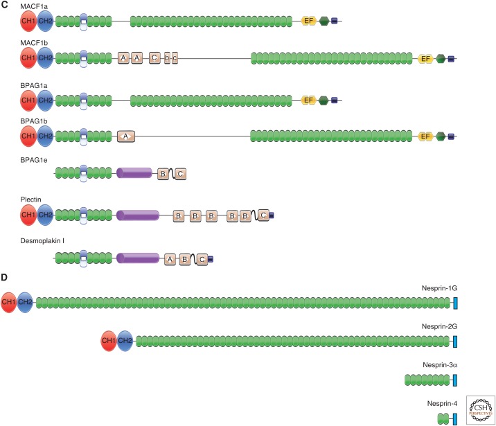
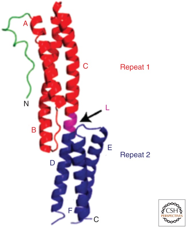
This review discusses the spectrin superfamily of proteins that function to connect cytoskeletal elements to each other, the cell membrane, and the nucleus. The signature domain is the spectrin repeat, a 106-122-amino-acid segment comprising three α-helices. α-actinin is considered to be the ancestral protein and functions to cross-link actin filaments. It then evolved to generate spectrin and dystrophin that function to link the actin cytoskeleton to the cell membrane, as well as the spectraplakins and plakins that link cytoskeletal elements to each other and to junctional complexes. A final class comprises the nesprins, which are able to bind to the nuclear membrane. This review discusses the domain organization of the various spectrin family members, their roles in protein-protein interactions, and their roles in disease, as determined from mutations, and it also describes the functional roles of the family members as determined from null phenotypes.


Figures

Figure 1.
Figure 1.
Schematic diagrams of the spectrin superfamily of cytoskeletal linker proteins. (A) α-actinin, spectrin, and dystrophin/utrophin. α-actinin is shown as an antiparallel dimer. The two actin-binding domains (ABDs) CH1 and CH2 allow the cross-linking of actin filaments. Spectrin is shown as a dimer of α- and β-spectrin, with α-spectrin shown below β-spectrin in an antiparallel orientation (from carboxyl to amino terminus). A tetramer can be formed by a second antiparallel dimer in the opposite direction, with the amino terminus of α-spectrin interacting with the carboxyl terminus of β-spectrin (Broderick and Winder 2005). The resulting tetramer resembles α-actinin, but has many more spectrin repeats (SRs). Dystrophin is shown as a monomer. It can interact with actin through the ABD, but has many more interaction domains, including a second ABD in the last two SRs. Utrophin is similar to dystrophin, except that the last two SRs are missing. The domain labeled “ZZ” is the zinc-finger domain. (B) Invertebrate spectraplakins. The two splice forms of the two known invertebrate spectraplakins are shown. Shots I and II are the Drosophila spectraplakins, and VAB-10a and VAB-10b are the Caenorhabditis elegans spectraplakins. The identity of the various domains is given in the key. (C) Vertebrate spectraplakins and plakins. The major “a” and “b” splice forms of microtubule-actin cross-linking factor 1 (MACF1) and bullous pemphigoid antigen 1 (BPAG1) are shown. MACF1a and MACF1b have domain structures similar to those of BPAG1a and BPAG1b, respectively. However, a third splice form of BPAG1—BPAG1e—only shares the so-called plakin domain in common with the other two isoforms. BPAG1e can dimerize through its coiled-coil rod. Plectin and desmoplakin, along with BPAG1e, are considered “plakins” rather than “spectraplakins,” although the plakin domain actually consists of SRs. (D) Nesprins. The four major isoforms of the nesprins are shown. There are some unusual SRs in the two giant isoforms, but, for simplicity, they are shown here as standard SRs.
Figure 1.
Figure 1.
Schematic diagrams of the spectrin superfamily of cytoskeletal linker proteins. (A) α-actinin, spectrin, and dystrophin/utrophin. α-actinin is shown as an antiparallel dimer. The two actin-binding domains (ABDs) CH1 and CH2 allow the cross-linking of actin filaments. Spectrin is shown as a dimer of α- and β-spectrin, with α-spectrin shown below β-spectrin in an antiparallel orientation (from carboxyl to amino terminus). A tetramer can be formed by a second antiparallel dimer in the opposite direction, with the amino terminus of α-spectrin interacting with the carboxyl terminus of β-spectrin (Broderick and Winder 2005). The resulting tetramer resembles α-actinin, but has many more spectrin repeats (SRs). Dystrophin is shown as a monomer. It can interact with actin through the ABD, but has many more interaction domains, including a second ABD in the last two SRs. Utrophin is similar to dystrophin, except that the last two SRs are missing. The domain labeled “ZZ” is the zinc-finger domain. (B) Invertebrate spectraplakins. The two splice forms of the two known invertebrate spectraplakins are shown. Shots I and II are the Drosophila spectraplakins, and VAB-10a and VAB-10b are the Caenorhabditis elegans spectraplakins. The identity of the various domains is given in the key. (C) Vertebrate spectraplakins and plakins. The major “a” and “b” splice forms of microtubule-actin cross-linking factor 1 (MACF1) and bullous pemphigoid antigen 1 (BPAG1) are shown. MACF1a and MACF1b have domain structures similar to those of BPAG1a and BPAG1b, respectively. However, a third splice form of BPAG1—BPAG1e—only shares the so-called plakin domain in common with the other two isoforms. BPAG1e can dimerize through its coiled-coil rod. Plectin and desmoplakin, along with BPAG1e, are considered “plakins” rather than “spectraplakins,” although the plakin domain actually consists of SRs. (D) Nesprins. The four major isoforms of the nesprins are shown. There are some unusual SRs in the two giant isoforms, but, for simplicity, they are shown here as standard SRs.
Figure 2.
Figure 2.
Ribbon diagram illustrating the structure of two spectrin repeats (SRs) in the plakin domain of bullous pemphigoid antigen 1 (BPAG1/dystonin), which is a member of the spectrin superfamily. Depicted is a loop-like region (green) at the far amino-terminal end, followed by a pair of SRs 1 and 2 (red and blue, respectively) arranged in tandem and connected by a linker region (L, purple) that is also helical in nature. Also labeled are the component helices A–C and D–F. (Reprinted from Jefferson et al. 2004.)
Figure 3.
Figure 3.
Depiction of the structure of the LINC (linker of nucleoskeleton and cytoskeleton) complex that bridges the cytoskeleton and nucleoskeleton. The complex comprises nesprin proteins (“KASH proteins”) in the outer nuclear membrane (ONM) and SUN proteins in the inner nuclear membrane (INM). Extending into the cytoplasm are extensions of the nesprins that are of variable size and interact with different cytoskeletal elements. The SUN proteins anchor the LINC complex to the nucleoskeleton through their nucleoplasmic domains by means of interaction with the nuclear lamina, as well as with chromosome-binding proteins and probably other anchoring proteins. (Reprinted from Chang et al. 2015.)

References

    1. Albrecht LV, Zheng L, Shabanowitz J, Purevjav E, Towbin JA, Hunt DF, Green KJ. 2015. GSK3- and PRMT-1 dependent modification of desmoplakin control desmoplakin-cytoskeleton dynamics. J Cell Biol 208: 597–612. - PMC - PubMed
    1. Andra K, Lassmann H, Bittner R, Shorny S, Fässler R, Probst F, Wiche G. 1997. Targeted inactivation of plectin reveals essential function in maintaining the integrity of skin, muscle and heart cytoarchitecture. Genes Dev 11: 3143–3156. - PMC - PubMed
    1. Bosher JM, Hahn BS, Legouis R, Sookhareea S, Weimer RM, Gansmuller A, Chisholm AD, Rose AM, Bessereau JL, Labouesse M. 2003. The Caenorhabditis elegans vab-10 spectraplakin isoforms protect the epidermis against internal and external forces. J Cell Biol 161: 757–768. - PMC - PubMed
    1. Broderick MJ, Winder SJ. 2002. Towards a complete atomic structure of spectrin family proteins. J Struct Biol 137: 184–193. - PubMed
    1. Broderick MJ, Winder SJ. 2005. Spectrin, α-actinin, and dystrophin. Adv Protein Chem 70: 203–246. - PubMed

LinkOut - more resources