Comparative Study
. 2016 Dec;68(6):1415-1423.
doi: 10.1161/HYPERTENSIONAHA.116.07850. Epub 2016 Oct 3.

Resting Afferent Renal Nerve Discharge and Renal Inflammation: Elucidating the Role of Afferent and Efferent Renal Nerves in Deoxycorticosterone Acetate Salt Hypertension

Affiliations
Comparative Study

Resting Afferent Renal Nerve Discharge and Renal Inflammation: Elucidating the Role of Afferent and Efferent Renal Nerves in Deoxycorticosterone Acetate Salt Hypertension

Christopher T Banek et al. Hypertension. 2016 Dec.

Abstract

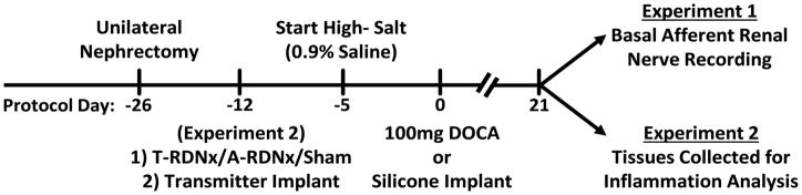
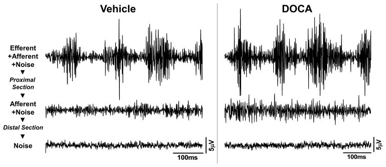
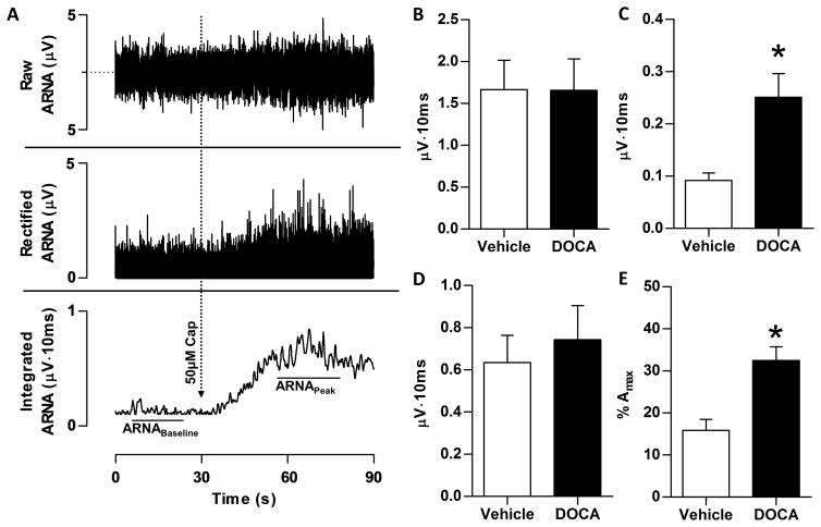
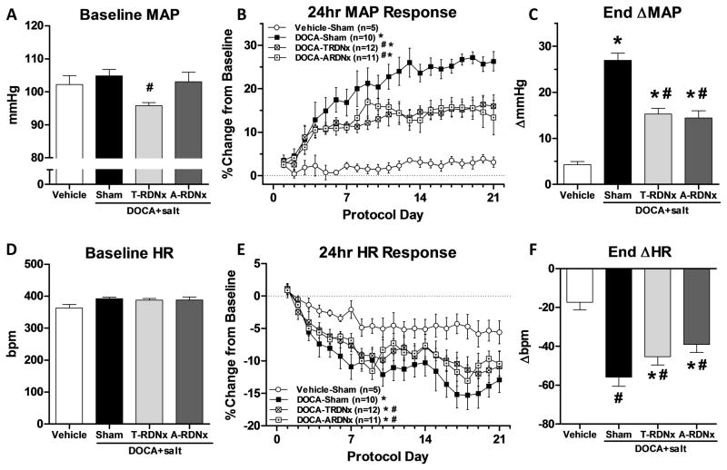
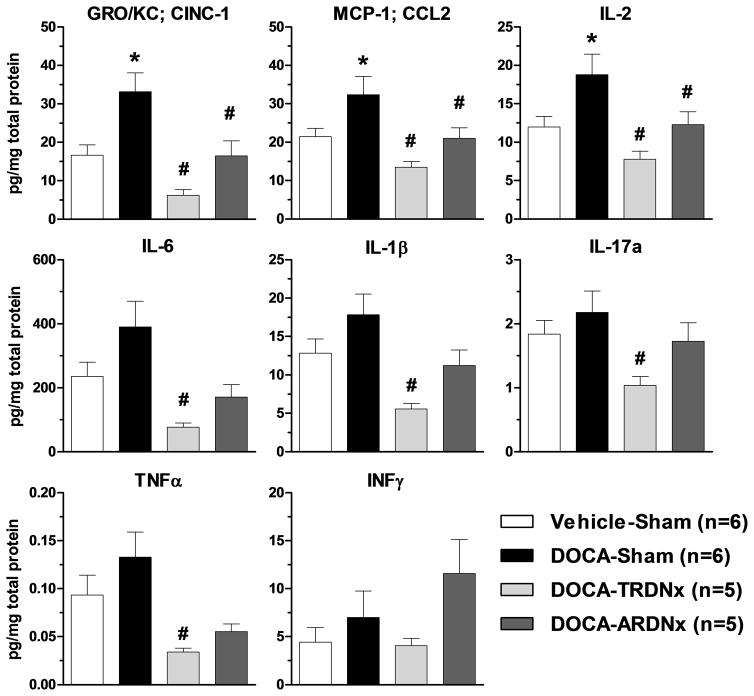
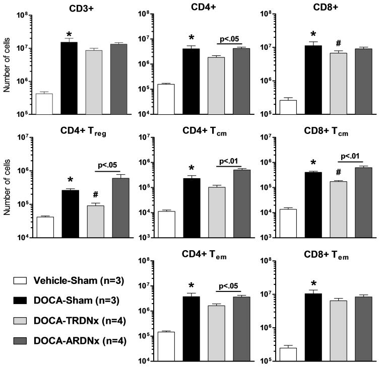
Renal sympathetic denervation (RDNx) has emerged as a novel therapy for hypertension; however, the therapeutic mechanisms remain unclear. Efferent renal sympathetic nerve activity has recently been implicated in trafficking renal inflammatory immune cells and inflammatory chemokine and cytokine release. Several of these inflammatory mediators are known to activate or sensitize afferent nerves. This study aimed to elucidate the roles of efferent and afferent renal nerves in renal inflammation and hypertension in the deoxycorticosterone acetate (DOCA) salt rat model. Uninephrectomized male Sprague-Dawley rats (275-300 g) underwent afferent-selective RDNx (n=10), total RDNx (n=10), or Sham (n=10) and were instrumented for the measurement of mean arterial pressure and heart rate by radiotelemetry. Rats received 100-mg DOCA (SC) and 0.9% saline for 21 days. Resting afferent renal nerve activity in DOCA and vehicle animals was measured after the treatment protocol. Renal tissue inflammation was assessed by renal cytokine content and T-cell infiltration and activation. Resting afferent renal nerve activity, expressed as a percent of peak afferent nerve activity, was substantially increased in DOCA than in vehicle (35.8±4.4 versus 15.3±2.8 %Amax). The DOCA-Sham hypertension (132±12 mm Hg) was attenuated by ≈50% in both total RDNx (111±8 mm Hg) and afferent-selective RDNx (117±5 mm Hg) groups. Renal inflammation induced by DOCA salt was attenuated by total RDNx and unaffected by afferent-selective RDNx. These data suggest that afferent renal nerve activity may mediate the hypertensive response to DOCA salt, but inflammation may be mediated primarily by efferent renal sympathetic nerve activity. Also, resting afferent renal nerve activity is elevated in DOCA salt rats, which may highlight a crucial neural mechanism in the development and maintenance of hypertension.

Keywords: afferent neurons; arterial pressure; denervation; deoxycorticosterone acetate; hypertension; inflammation.


Figures

Figure 1
Figure 1
The DOCA-salt protocol timeline for experiments 1 and 2.
Figure 2
Figure 2
Examples of original nerve tracings for total renal nerve activity (efferent+afferent+noise), afferent(+noise), and background noise from a vehicle and DOCA rat. Afferent signal was recorded after proximal sectioning of the isolated renal nerve. Background noise was recorded following a distal sectioning of the isolated nerve. Scale bars: 100ms (x-axis); 5μV (y-axis).
Figure 3
Figure 3
Following DOCA-salt treatment, resting afferent renal nerve activity (ARNA) was measured in anesthetized rats. Panel A: A sample tracing of raw (top), rectified (middle), and integrated (bottom) nerve activity from a Vehicle-Sham animal is depicted, in which ARNA was recorded before and during the response to intrapelvic administration of 50μM capsaicin to establish the peak level of afferent renal nerve discharge. ARNA was quantified from the integrated (∫ARNA) signal. Panel B: Raw integrated signal of resting total renal nerve activity is no difference between Vehicle and DOCA rats. Panel C: Following proximal sectioning of the renal nerve, signal recorded represents only ∫ARNA. The resting ∫ARNA was elevated in DOCA versus Vehicle (0.24±0.04 versus 0.10±0.01 μV·sec). Panel D: Peak afferent nerve response to intrapelvic perfusion of 50μM capsaicin was no different between Vehicle and DOCA. Panel E: Basal ARNA, expressed as a percentage of peak ARNA response to intrapelvic capsaicin (%Amax). After normalization, resting activity remained increased in DOCA-salt rats compared Vehicle (32.0±5.7 versus 13.8±2.7%Amax). All data presented as mean±SEM (n=10/group). *p<.05 versus Vehicle-Sham.
Figure 4
Figure 4
Panel A: Baseline mean arterial pressure (MAP) prior to DOCA treatment, was lower in T-RDNx group versus DOCA-Sham (*96±1 versus 105±2mmHg) rats and A-RDNx had no effect (103±4mmHg). Panel B: The percent increase from baseline MAP in DOCA-Sham rats was attenuated equally by T-RDNx and A-RDNx. Panel C: The end ΔMAP was averaged from the final three days of treatment (days 19–21) in the four treatment groups. The ΔMAP DOCA-Sham (27±2mmHg) was halved by T-RDNx and A-RDNx (15±1 and 14±2mmHg). Panel D: No differences in baseline HR was observed. Panel E: The percent change in heart rate from baseline was reduced in DOCA-Sham compared to Vehicle-Sham rats, and both T-RDNx and A-RDNx mitigated this effect. Panel F: End ΔHR averaged of the final three days of treatment was decreased in DOCA-sham versus Vehicle-Sham (−56±5 versus −17±4bpm), and T-RDNx and A-RDNx attenuated this response (−45±4 versus −9±4bpm). All data presented as mean±SEM. *p<.05 versus Vehicle-Sham; #p<.05 versus DOCA-Sham.
Figure 5
Figure 5
Renal cortical and medullary (mixed) tissue inflammatory marker content was measured by Luminex multiplex immunoassay. Several pro-inflammatory markers (GRO/KC, MCP-1, IL-2, and IL-6) were increased (*p<.05) in DOCA-Sham rats versus Vehicle-Sham. T-RDNx greatly decreased (#p<.05) the levels of GRO/KC, MCP-1, IL-1β, IL-2, IL-6, IL-17a, and TNFα versus DOCA-Sham. A-RDNx attenuated fewer pro-inflammatory cytokines (GRO/KC, MCP-1, IL-2) versus DOCA-Sham. All data presented as mean±SEM. *p<.05 versus Vehicle-Sham; #p<.05 versus DOCA-Sham.
Figure 6
Figure 6
Renal T-cell infiltration and activation were assessed by flow cytometry. Nearly all T-cells subtypes were elevated with DOCA-salt treatment. Total renal nerve ablation (T-RDNx) tended to lower renal T-cell infiltration and activation versus DOCA-Sham. Compared to afferent-targeted RDNx (A-RDNx), CD4+ central memory (Tcm), effector memory (Tem), and regulatory (Treg) were lower in T-RDNx animals. All data presented as mean±SEM. *p<.05 versus Vehicle-Sham; #p<.05 versus DOCA-Sham.

References

    1. Mozaffarian D, Benjamin EJ, Go AS, et al. Heart disease and stroke statistics-2016 update: A report from the american heart association. Circulation. 2016;133:e38–e360. - PubMed
    1. Kearney PM, Whelton M, Reynolds K, Muntner P, Whelton PK, He J. Global burden of hypertension: Analysis of worldwide data. Lancet. 2005;365:217–223. - PubMed
    1. Davis MI, Filion KB, Zhang D, Eisenberg MJ, Afilalo J, Schiffrin EL, Joyal D. Effectiveness of renal denervation therapy for resistant hypertension: A systematic review and meta-analysis. J Am Coll Cardiol. 2013;62:231–241. - PubMed
    1. DiBona GF, Esler M. Translational medicine: The antihypertensive effect of renal denervation. Am J Physiol Regul Integr Comp Physiol. 2010;298:R245–253. - PubMed
    1. Krum H, Schlaich M, Whitbourn R, Sobotka PA, Sadowski J, Bartus K, Kapelak B, Walton A, Sievert H, Thambar S, Abraham WT, Esler M. Catheter-based renal sympathetic denervation for resistant hypertension: A multicentre safety and proof-of-principle cohort study. Lancet. 2009;373:1275–1281. - PubMed

Publication types

Substances