. 2016 Nov 18;198(24):3345-3354.
doi: 10.1128/JB.00625-16. Print 2016 Dec 15.

Aag Hypoxanthine-DNA Glycosylase Is Synthesized in the Forespore Compartment and Involved in Counteracting the Genotoxic and Mutagenic Effects of Hypoxanthine and Alkylated Bases in DNA during Bacillus subtilis Sporulation

Affiliations

Aag Hypoxanthine-DNA Glycosylase Is Synthesized in the Forespore Compartment and Involved in Counteracting the Genotoxic and Mutagenic Effects of Hypoxanthine and Alkylated Bases in DNA during Bacillus subtilis Sporulation

Víctor M Ayala-García et al. J Bacteriol. .

Abstract

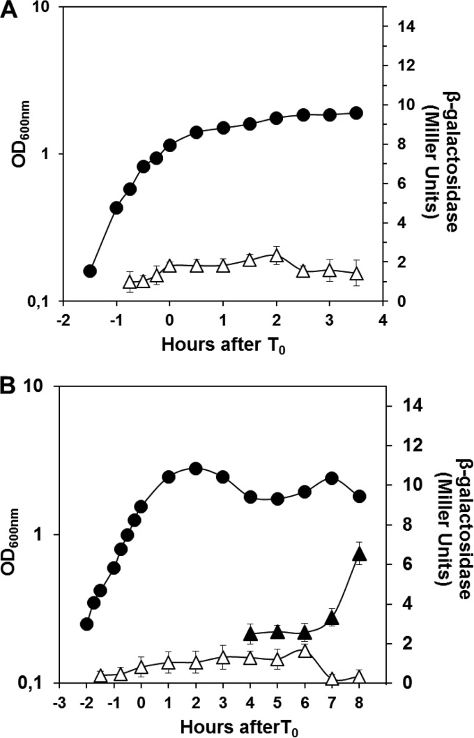
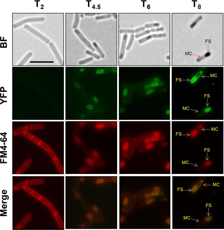
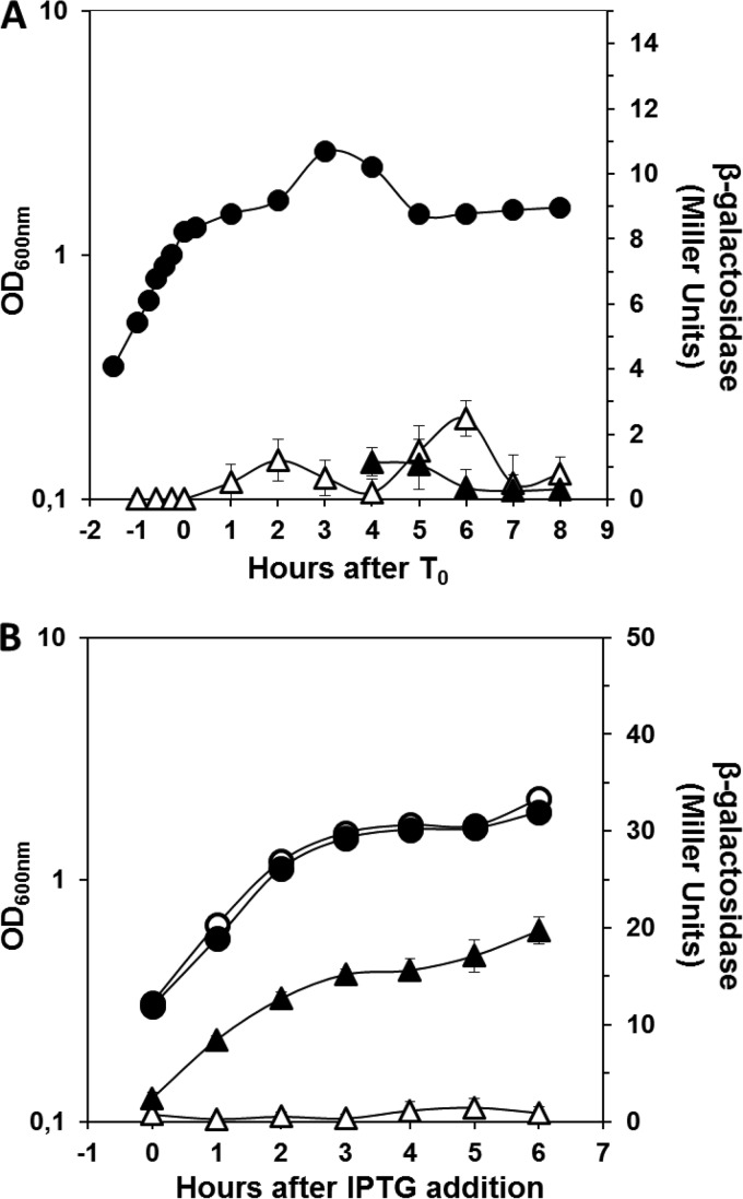
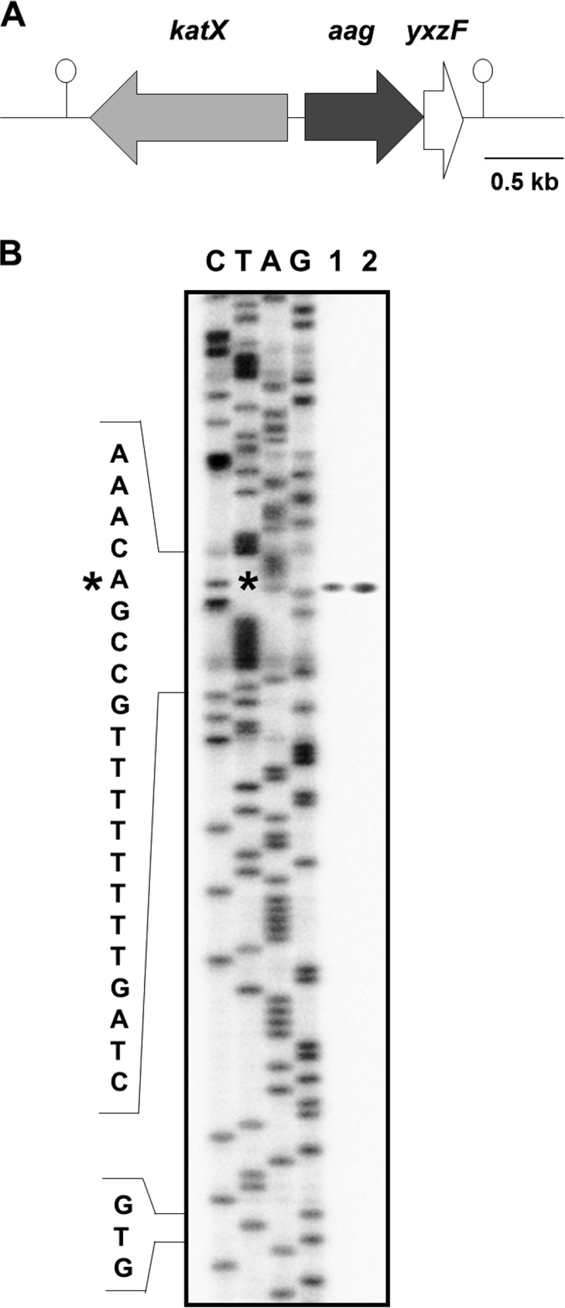
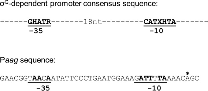
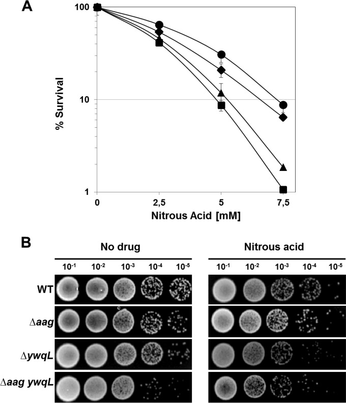
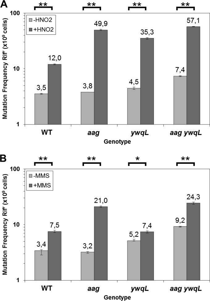
Aag from Bacillus subtilis has been implicated in in vitro removal of hypoxanthine and alkylated bases from DNA. The regulation of expression of aag in B. subtilis and the resistance to genotoxic agents and mutagenic properties of an Aag-deficient strain were studied here. A strain with a transcriptional aag-lacZ fusion expressed low levels of β-galactosidase during growth and early sporulation but exhibited increased transcription during late stages of this developmental process. Notably, aag-lacZ expression was higher inside the forespore than in the mother cell compartment, and this expression was abolished in a sigG-deficient background, suggesting a forespore-specific mechanism of aag transcription. Two additional findings supported this suggestion: (i) expression of an aag-yfp fusion was observed in the forespore, and (ii) in vivo mapping of the aag transcription start site revealed the existence of upstream regulatory sequences possessing homology to σG-dependent promoters. In comparison with the wild-type strain, disruption of aag significantly reduced survival of sporulating B. subtilis cells following nitrous acid or methyl methanesulfonate treatments, and the Rifr mutation frequency was significantly increased in an aag strain. These results suggest that Aag protects the genome of developing B. subtilis sporangia from the cytotoxic and genotoxic effects of base deamination and alkylation.

Importance: In this study, evidence is presented revealing that aag, encoding a DNA glycosylase implicated in processing of hypoxanthine and alkylated DNA bases, exhibits a forespore-specific pattern of gene expression during B. subtilis sporulation. Consistent with this spatiotemporal mode of expression, Aag was found to protect the sporulating cells of this microorganism from the noxious and mutagenic effects of base deamination and alkylation.


Figures

FIG 1
FIG 1
Expression of aag during B. subtilis growth and sporulation. (A) β-Galactosidase activity from an aag-lacZ fusion during vegetative growth. B. subtilis strain PERM1246 harboring an aag-lacZ fusion was grown in PAB medium and the OD600 measured (●). Cells were collected at various times, and the β-galactosidase activity of growing cells (△) was determined as described in Materials and Methods. Values for β-galactosidase specific activity are the averages of values from three independent experiments ± standard deviations (SD). (B) β-Galactosidase specific activity from an aag-lacZ fusion during sporulation. B. subtilis strain PERM1246 harboring an aag-lacZ fusion was induced to sporulate in DSM and the OD600 measured (●). Time zero of sporulation (T0) is defined as the time when log-phase cell growth stops. Cells were collected at various times, and β-galactosidase assays were conducted in both mother cell (△) and forespore (▲) fractions as described in Materials and Methods. Values for β-galactosidase specific activity are the averages of values from three independent experiments ± SD.
FIG 2
FIG 2
Localization of Aag-YFP during B. subtilis sporulation. B. subtilis strain PERM1286 harboring a translational in-frame aag-yfp fusion was grown and sporulated in DSM. At 2, 4.5, 6, and 8 h after the onset of sporulation, sporulating cells were analyzed by bright-field (BF) and fluorescence (YFP and FM4-64) microscopy as described in Materials and Methods. Overlain images of YFP and FM4-64 at each time point are depicted as merge. MC, mother cell; FS, forespore compartments. Scale bar, 5 μM.
FIG 3
FIG 3
σG dependence of aag-lacZ expression. (A) β-Galactosidase activity of aag-lacZ during sporulation of the ΔsigG strain. B. subtilis strain PERM1375 harboring an aag-lacZ fusion in a ΔsigG genetic background was sporulated in DSM and the OD600 measured (●). Cells were collected at various times, and β-galactosidase activity was assayed in mother cell (△) and forespore (▲) fractions as described in Materials and Methods. Values for β-galactosidase specific activity are the averages of values from three independent experiments ± SD. (B) β-Galactosidase activity from an aag-lacZ fusion induced by overexpression of sigG during vegetative growth. B. subtilis strains PERM1375 and PERM1374 harboring an aag-lacZ fusion in a ΔsigG genetic background containing either a Pspac::sigG gene fusion (●) or the empty vector (○) were grown in PAB medium and induced with IPTG (1 mM) when an OD600 of 0.5 was reached. The OD600s of cultures were measured (● and ○), cells were collected at various times, and β-galactosidase assays from cells that were (▲) or were not (△) complemented with sigG were conducted as described in Materials and Methods. Values for β-galactosidase specific activity are the averages of values from three independent experiments ± SD.
FIG 4
FIG 4
Genetic map of the aag region and mapping of the aag promoter. (A) The B. subtilis chromosomal region around aag. The katX gene is divergently transcribed upstream of aag, and aag and yxzF may comprise a bicistronic operon. Hairpin structures denote putative transcriptional terminators. The drawing is to scale. (B) Primer extension analysis to identify the 5′ end of aag mRNA. Total RNA was isolated from T6 sporulating cells of B. subtilis strain 168 (wild type) grown in DSM (lane 1) or vegetative cells of strain PERM1375 grown in PAB medium, induced with IPTG, and harvested 3 h after IPTG addition (lane 2). Primer extension was performed as described in Materials and Methods. The asterisk indicates the position of the primer extension product relative to the DNA sequencing ladder (lanes C, T, A, and G). The position of the mapped 5′ end of aag mRNA is denoted with an asterisk. “GTG” indicates the position of the aag translation start codon.
FIG 5
FIG 5
Comparison of the consensus σG (39) promoter sequence with the putative promoter sequences (Paag) upstream of the aag ORF. Perfect matches are in bold. The position of the aag transcription start site is denoted with an asterisk. Abbreviations: H, A or C; R, A or G; and X, A or T.
FIG 6
FIG 6
Sensitivity of sporulating cells to HNO2. (A) Survival of B. subtilis wild-type (●), aag (◆), ywqL (▲), and aag ywqL (■) strains following HNO2 treatment. Cells from various strains were sporulated in liquid DSM and treated with different concentrations of HNO2 at 4.5 h after the onset of sporulation, and cell viability was determined as described in Materials and Methods. Results are expressed as averages ± SD from at least three independent experiments. (B) Resistance of B. subtilis wild-type (WT), aag, ywqL, and aag ywqL strains after exposure to HNO2. Cells from all strains were sporulated in liquid DSM, treated with the LD50 of HNO2 (calculated from the dose-response curve in panel A for the wild-type strain) at 4.5 h after the onset of sporulation, spot plated, grown, and photographed as described in Materials and Methods.
FIG 7
FIG 7
Sensitivity of sporulating cells to MMS. (A) Killing of B. subtilis wild-type (●), aag (◆), ywqL (▲), and aag ywqL (■) strains by MMS. Cells from all strains were sporulated in liquid DSM and treated with different concentrations of MMS at 4.5 h after the onset of sporulation, and cell viability was determined as described in Materials and Methods. Results are expressed as averages ± SD from at least three independent experiments. (B) Resistance of B. subtilis wild-type (WT), aag, ywqL, and aag ywqL strains after exposure to MMS. Cells from all strains were sporulated in liquid DSM, treated with the LD50 of MMS (calculated from the dose-response curve in panel A for the wild-type strain) at 4.5 h after the onset of sporulation, spot plated, grown, and photographed as described in Materials and Methods.
FIG 8
FIG 8
Frequencies of mutation to Rifr. Cells of B. subtilis wild-type, aag, ywqL, and aag ywqL strains were sporulated in liquid DSM and treated with the LD50 for each strain of either HNO2 (A) or MMS (B) at 4.5 h after the onset of sporulation. The levels of Rifr cells with (+) or without (−) treatment exposure were determined. Each bar represents the mean of data collected from three independent experiments, and error bars represent the standard error of the mean (SEM). *, P < 0.05; **, P < 0.01 (by the Mann-Whitney U test).

References

    1. Friedberg EC, Walker GC, Siede W, Wood RD, Schultz RA, Ellenberger T. 2006. DNA repair and mutagenesis, 2nd ed American Society for Microbiology, Washington, DC.
    1. Dalhus B, Laerdahl JK, Backe PH, Bjørås M. 2009. DNA base repair—recognition and initiation of catalysis. FEMS Microbiol Rev 33:1044–1078. doi:10.1111/j.1574-6976.2009.00188.x. - DOI - PubMed
    1. Yonekura SI, Nakamura N, Yonei S, Zhang-Akiyama QM. 2009. Generation, biological consequences and repair mechanisms of cytosine deamination in DNA. J Radiat Res 50:19–26. doi:10.1269/jrr.08080. - DOI - PubMed
    1. Lindahl T. 1993. Instability and decay of the primary structure of DNA. Nature 362:709–715. doi:10.1038/362709a0. - DOI - PubMed
    1. Barnes DE, Lindahl T. 2004. Repair and genetic consequences of endogenous DNA base damage in mammalian cells. Annu Rev Genet 38:445–476. doi:10.1146/annurev.genet.38.072902.092448. - DOI - PubMed

Publication types

MeSH terms

LinkOut - more resources