. 2016 Nov 18;198(24):3318-3328.
doi: 10.1128/JB.00548-16. Print 2016 Dec 15.

Intracellular d-Serine Accumulation Promotes Genetic Diversity via Modulated Induction of RecA in Enterohemorrhagic Escherichia coli

Affiliations

Intracellular d-Serine Accumulation Promotes Genetic Diversity via Modulated Induction of RecA in Enterohemorrhagic Escherichia coli

James P R Connolly et al. J Bacteriol. .

Abstract

We recently discovered that exposure of enterohemorrhagic Escherichia coli (EHEC) to d-serine resulted in accumulation of this unusual amino acid, induction of the SOS regulon, and downregulation of the type III secretion system that is essential for efficient colonization of the host. Here, we have investigated the physiological relevance of this elevated SOS response, which is of particular interest given the presence of Stx toxin-carrying lysogenic prophages on the EHEC chromosome that are activated during the SOS response. We found that RecA elevation in response to d-serine, while being significant, was heterogeneous and not capable of activating stx expression or stx phage transduction to a nonlysogenic recipient. This "SOS-like response" was, however, capable of increasing the mutation frequency associated with low-level RecA activity, thus promoting genetic diversity. Furthermore, this response was entirely dependent on RecA and enhanced in the presence of a DNA-damaging agent, indicating a functional SOS response, but did not result in observable cleavage of the LexA repressor alone, indicating a controlled mechanism of induction. This work demonstrates that environmental factors not usually associated with DNA damage are capable of promoting an SOS-like response. We propose that this modulated induction of RecA allows EHEC to adapt to environmental insults such as d-serine while avoiding unwanted phage-induced lysis.

Importance: The SOS response is a global stress network that is triggered by the presence of DNA damage due to breakage or stalled replication forks. Activation of the SOS response can trigger the replication of lytic bacteriophages and promote genetic diversification through error-prone polymerases. We have demonstrated that the host-associated metabolite d-serine contributes to Escherichia coli niche specification and accumulates inside cells that cannot catabolize it. This results in a modulated activation of the SOS antirepressor RecA that is insufficient to trigger lytic bacteriophage but capable of increasing the SOS-associated mutation frequency. These findings describe how relevant signals not normally associated with DNA damage can hijack the SOS response, promoting diversity as E. coli strains adapt while avoiding unwanted phage lysis.


Figures

FIG 1
FIG 1
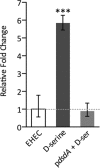
Induction of recA transcription upon intracellular d-serine accumulation. qRT-PCR analysis of relative recA transcription with mRNA from EHEC cultured to mid-exponential phase in MEM-HEPES alone and supplemented with 1 mM d-serine. As a control, recA levels from an EHEC strain transformed with pdsdA (encoding d-serine deaminase from UPEC) that does not accumulate d-serine were also analyzed. The broken line indicates the baseline wild-type recA level. ***, P ≤ 0.001 (calculated from three independent biological replicates). Error bars indicate standard deviations.
FIG 2
FIG 2
d-Serine does not induce lytic bacteriophage and stx expression in EHEC. Growth of EHEC was profiled in MEM-HEPES supplemented with 1, 0.2, or 0.02 mM d-serine (A) and 2, 0.5, or 0.05 μg/ml MMC (B). The time point of SOS induction by d-serine or MMC is indicated by an arrow. A sharp drop in OD600 is indicative of phage-induced lysis. (C) stx-GFP reporter expression in response to d-serine and MMC was analyzed. Relative fluorescence units (RFU) were measured at various time points postinduction and compared to those of uninduced EHEC. **, P ≤ 0.01; ***, P ≤ 0.001 (calculated from three independent biological replicates). (D) Quantification of the number of phage transduction events in response to a d-serine- or MMC-induced SOS response in EHEC. Error bars represent the SEM calculated from at least three biological replicates. The concentrations of d-serine and MMC used are indicated individually in each panel.
FIG 3
FIG 3
Increased SOS-like mutation frequency after d-serine accumulation. The emergence of resistance to rifampin (Rifr) in EHEC after SOS induction by 1 mM d-serine and 0.05 μg/ml MMC was assayed. EHEC cell cultures were harvested and plated on rifampin-containing LB plates in exponential phase at 2 h postinduction (A) and in stationary phase at 8 h postinduction (B). The dotted line indicates the background level of spontaneous Rifr emergence in uninduced EHEC cells, to which all other conditions were compared. The Rifr frequency after each incubation is relative to the number of CFU present in each culture. As a control, EHEC/pdsdA, which does not accumulate d-serine, was also assayed for the emergence of Rifr. Error bars represent the SEM calculated from at least five biological replicates. *, P ≤ 0.05 (compared to the wild-type control).
FIG 4
FIG 4
Modulated expression of recA induced by d-serine accumulation. (A) recA-GFP reporter expression was analyzed in response to d-serine and MMC. Relative fluorescence units (RFU) were measured at various stages of growth postinduction and compared to those of uninduced EHEC at all of the time points tested. The concentrations of d-serine or MMC used are indicated individually in each panel. Error bars represent the SEM. *, P ≤ 0.05; **, P ≤ 0.01; ***, P ≤ 0.001 (calculated from three independent biological replicates). (B) Fluorescence microscopy analysis of EHEC/precA-GFP cell populations left untreated or treated with 1 mM d-serine or 2.0 μg/ml MMC. Bacterial cells were imaged by phase-contrast microscopy, and recA induction was determined by detection of GFP-expressing cells. The merged channel indicates the level of recA-GFP induction per cell. Representative images are displayed for each condition, and data are summarized below as the percentage of the population expressing enhanced recA-GFP activity (green bars, cells induced for recA-GFP expression; gray bars, cells uninduced for recA-GFP expression). Experiments were performed in triplicate, and the results are shown as the mean ± the SEM. WT, wild type. (C) Immunoblot analysis of RecA, LexA, and DnaK levels in whole-cell lysates of EHEC cultured in MEM-HEPES with or without SOS induction. An EHEC ΔrecA mutant was used as a negative control for SOS induction. The concentrations of d-serine and MMC used to induce the SOS response are indicated above the immunoblots. DnaK levels were used to assess equal protein loading of samples. Experiments were performed at least in triplicate to confirm the results.
FIG 5
FIG 5
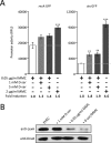
d-Serine (d-ser) accumulation induces a functional SOS-like response in EHEC. (A) The levels of recA-GFP (left) and stx-GFP (right) reporter expression were analyzed in cell cultures treated with 0.05 or 2.0 μg/ml MMC. The levels of reporter activity in response to a combination of a low MMC concentration supplemented with either 1 or 5 mM d-serine were also analyzed and compared to that of a low MMC concentration. Relative fluorescence units (RFU) were measured at 3 h postinduction (OD600, ∼0.8). The concentrations and combinations of d-serine and MMC used are indicated at the bottom, where a plus sign means present and a minus sign means absent. The broken line indicates the level of recA or stx expression in response to a low subinhibitory MMC concentration. Error bars represent the SEM. **, P ≤ 0.01; ***, P ≤ 0.001 (calculated from three independent biological replicates). The enhanced SOS response is also represented as the fold change in recA or stx expression indicated at the bottom. (B) Immunoblot analysis of LexA and DnaK levels in whole-cell lysates of EHEC induced for an SOS response with d-serine, MMC, or both. The concentration of d-serine or MMC used to induce the SOS response is indicated above the immunoblots. DnaK levels were used to assess equal protein loading of samples. Experiments were performed at least in triplicate to confirm the results.

Similar articles

Cited by

References

    1. Baharoglu Z, Mazel D. 2014. SOS, the formidable strategy of bacteria against aggressions. FEMS Microbiol Rev 38:1126–1145. doi:10.1111/1574-6976.12077. - DOI - PubMed
    1. Michel B. 2005. After 30 years of study, the bacterial SOS response still surprises us. PLoS Biol 3:e255. doi:10.1371/journal.pbio.0030255. - DOI - PMC - PubMed
    1. Fernández De Henestrosa AR, Ogi T, Aoyagi S, Chafin D, Hayes JJ, Ohmori H, Woodgate R. 2000. Identification of additional genes belonging to the LexA regulon in Escherichia coli. Mol Microbiol 35:1560–1572. - PubMed
    1. Courcelle J, Khodursky A, Peter B, Brown PO, Hanawalt PC. 2001. Comparative gene expression profiles following UV exposure in wild-type and SOS-deficient Escherichia coli. Genetics 158:41–64. - PMC - PubMed
    1. Wade JT, Reppas NB, Church GM, Struhl K. 2005. Genomic analysis of LexA binding reveals the permissive nature of the Escherichia coli genome and identifies unconventional target sites. Genes Dev 19:2619–2630. doi:10.1101/gad.1355605. - DOI - PMC - PubMed

MeSH terms

LinkOut - more resources