. 2016 Oct 18;113(42):11871-11876.
doi: 10.1073/pnas.1606857113. Epub 2016 Oct 3.

Enhancer priming by H3K4 methyltransferase MLL4 controls cell fate transition

Affiliations

Enhancer priming by H3K4 methyltransferase MLL4 controls cell fate transition

Chaochen Wang et al. Proc Natl Acad Sci U S A. .

Abstract

Transcriptional enhancers control cell-type-specific gene expression. Primed enhancers are marked by histone H3 lysine 4 (H3K4) mono/di-methylation (H3K4me1/2). Active enhancers are further marked by H3K27 acetylation (H3K27ac). Mixed-lineage leukemia 4 (MLL4/KMT2D) is a major enhancer H3K4me1/2 methyltransferase with functional redundancy with MLL3 (KMT2C). However, its role in cell fate maintenance and transition is poorly understood. Here, we show in mouse embryonic stem cells (ESCs) that MLL4 associates with, but is surprisingly dispensable for the maintenance of, active enhancers of cell-identity genes. As a result, MLL4 is dispensable for cell-identity gene expression and self-renewal in ESCs. In contrast, MLL4 is required for enhancer-binding of H3K27 acetyltransferase p300, enhancer activation, and induction of cell-identity genes during ESC differentiation. MLL4 protein, rather than MLL4-mediated H3K4 methylation, controls p300 recruitment to enhancers. We also show that, in somatic cells, MLL4 is dispensable for maintaining cell identity but essential for reprogramming into induced pluripotent stem cells. These results indicate that, although enhancer priming by MLL4 is dispensable for cell-identity maintenance, it controls cell fate transition by orchestrating p300-mediated enhancer activation.

Keywords: H3K4 methyltransferase; MLL4/KMT2D; cell fate transition; enhancer; p300.


Conflict of interest statement

The authors declare no conflict of interest.

Figures
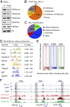
Fig. 1.
Fig. 1.
MLL4 associates with active enhancers on cell-identity genes in ESCs. (A) KO of the Mll4 gene in f/f (Mll3−/−;Mll4flox/flox) ESCs decreases global levels of H3K4me1/2. Histone extracts from f/f and Mll3/4 DKO ESCs were analyzed with immunoblotting using indicated antibodies. (B) Pie chart illustrating that MLL4+-binding sites are marked by H3K4me1 and H3K4me2 in ESCs. (C) MLL4 is enriched on AE regions in ESCs. Promoter: ±1 kb of transcription start site (TSS). AE: H3K4me1+;H3K27ac+ promoter-distal region. Primed enhancer: H3K4me1+;H3K27ac promoter-distal region. (D) Motif analysis of 5,918 MLL4-positive (MLL4+) AEs in ESCs. (E) MLL4 colocalizes with pluripotent TFs O/S/N (Oct4, Sox2, and Nanog) and H3K27 acetyltransferase p300 on AEs in ESCs. Density maps are shown for ChIP-Seq data at MLL4+ AEs. Color scale indicates ChIP-Seq signal in reads per million. (F) ChIP-Seq binding profiles (reads per million) of MLL4, p300, Oct4, and histone modifications on the ESC identity genes Nanog, Oct4 (Pou5f1), and Sox2. MLL4+ AEs are shaded in gray.
Fig. 2.
Fig. 2.
MLL4 is dispensable for maintaining ESC identity. (A and B) MLL4 is dispensable for maintaining ESC identity gene expression. (A) RNA-Seq was done in f/f and DKO ESCs. Expression levels of MLL4+ AE-associated genes are shown in the scatter plot. Twofold change threshold lines are indicated. Each dot represents one gene. MLL4-dependent and -independent genes are highlighted in red and blue, respectively. Note that MLL4 is required for Alpl expression in ESCs. (B) Expression fold changes of genes associated with MLL4+ and MLL4 TEs and SEs, respectively, were obtained by comparing DKO with f/f cells. (C and D) MLL4 is dispensable for ESC self-renewal. (C) Population doubling time of f/f and DKO cells. (D) Representative images of AP staining of ESC colonies. (E and F) MLL4 is dispensable for maintaining AEs on ESC identity genes. (E) Heat maps depicting fold changes of H3K4me1 and H3K27ac on MLL4+ AEs were obtained by comparing DKO with f/f cells. Enhancers, which show over threefold decreases of both H3K4me1 and H3K27ac levels in DKO cells, are defined as MLL4-dependent AEs. The rest are defined as MLL4-independent AEs. Selected genes associated with each group are shown on the right. (F) Average profiles of p300 around the center of MLL4-independent and -dependent AEs. The reduction of p300 binding on MLL4-dependent AEs is significantly larger than that on MLL4-independent ones (P value = 1.8E-19) (SI Appendix, SI Materials and Methods). (G) qRT-PCR analysis of expression of genes associated with MLL4-independent or MLL4-dependent AEs in f/f and DKO ESCs. (H) MLL4-independent and MLL4-dependent AEs on Nanog and Lefty1 gene loci, respectively, are shown. (Upper) ChIP-Seq binding profiles (reads per million) for MLL4, p300, Oct4, histone modifications, and mRNA-Seq signals in f/f and DKO cells. MLL4+ AEs are shaded in gray. (Lower) Interactions between MLL4+ AEs and promoters were determined by 3C assays in f/f and DKO ESCs. Enhancer–promoter interactions detected by Smc1 ChIA-PET were obtained from GSE57911 (12) and are shown above ChIP-Seq tracks. TSSs are indicated by red arrowheads.
Fig. 3.
Fig. 3.
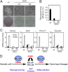
MLL4 is required for ESC differentiation. ESCs were induced to differentiate into EBs for 12 d. (A) Representative microscopic images of EBs at indicated time points. (B) qRT-PCR analysis of ESC markers (Nanog, Oct4), mesoderm markers (Brachyury, Wnt3), and endoderm markers (Gata4, Gata6) at indicated time points. (C) Representative histological images of teratomas derived from f/f and DKO ESCs. Three germ layers—ectoderm, mesoderm and endoderm—were observed in f/f teratomas, whereas cells in DKO teratomas remained at the primitive stage. A, adipose; C, cartilage; G, gland-like; M, muscle; NE, neural epithelium.
Fig. 4.
Fig. 4.
MLL4 is required for p300-mediated enhancer activation during ESC differentiation. f/f and DKO ESCs were induced to differentiate into EBs for 4 d, followed by ChIP-Seq of MLL4, p300, H3K4me1, H3K27ac, and RNA-Seq analyses. (A) Genomic distribution of MLL4-binding regions in D4 EBs. (B) Pie chart depicting that among MLL4+ AEs in D4 EBs derived from f/f ESCs. A total of 6,534 are de novo, whereas 9,704 are premarked with H3K27ac in ESCs. (C) Deletion of Mll4 in f/f ESCs prevents H3K27ac deposition and p300 recruitment on de novo AEs. Average profiles of H3K4me1, H3K27ac, and p300 around the center of premarked or de novo AEs are shown. The reduction of H3K27ac and p300 in DKO EBs on de novo AEs is significantly greater than that on premarked ones (P value = 4.4E-253 and 5.2E-97, respectively) (SI Appendix, SI Materials and Methods). (D) Box plot depicting the induction fold change of genes associated with premarked or de novo AEs. Only genes with a regulatory potential score over 2 are shown. P value was calculated using a t test. (E) ChIP-Seq binding profiles (reads per million) for MLL4, p300, H3K4me1, H3K27ac, and RNA-Seq signals on the Gata4 gene locus in ESCs and D4 EBs. MLL4+ de novo AEs are shaded in gray. TSS is indicated by a red arrowhead.
Fig. 5.
Fig. 5.
MLL4 is required for somatic cell reprogramming to iPSCs. Immortalized f/f and DKO MEFs were induced to reprogram for 20 d by infection with lentiviral vectors expressing TFs Oct4, Sox2, Klf4, and c-Myc (OSKM). (A) Representative images of reprogrammed cells at day 20. (Upper) Cell morphologies under microscope. (Lower) AP staining of dishes. (B) Reprogramming efficiencies of f/f MEFs and two DKO MEF cell lines. (C) qRT-PCR analysis of indicated genes in f/f and DKO cells before and after 20 d of reprogramming. (D) A model depicting the essential role of MLL3/MLL4 in ESC differentiation and self-renewal and somatic cell reprogramming. LDTFs, lineage-determining TFs.

References

    1. Heintzman ND, et al. Histone modifications at human enhancers reflect global cell-type-specific gene expression. Nature. 2009;459(7243):108–112. - PMC - PubMed
    1. Heinz S, Romanoski CE, Benner C, Glass CK. The selection and function of cell type-specific enhancers. Nat Rev Mol Cell Biol. 2015;16(3):144–154. - PMC - PubMed
    1. Calo E, Wysocka J. Modification of enhancer chromatin: What, how, and why? Mol Cell. 2013;49(5):825–837. - PMC - PubMed
    1. Whyte WA, et al. Master transcription factors and mediator establish super-enhancers at key cell identity genes. Cell. 2013;153(2):307–319. - PMC - PubMed
    1. Keller G. Embryonic stem cell differentiation: Emergence of a new era in biology and medicine. Genes Dev. 2005;19(10):1129–1155. - PubMed

Publication types

MeSH terms