. 2016 Oct 4:7:12907.
doi: 10.1038/ncomms12907.

Deubiquitinase activity is required for the proteasomal degradation of misfolded cytosolic proteins upon heat-stress

Affiliations

Deubiquitinase activity is required for the proteasomal degradation of misfolded cytosolic proteins upon heat-stress

Nancy N Fang et al. Nat Commun. .

Abstract

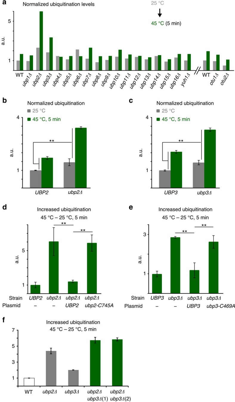
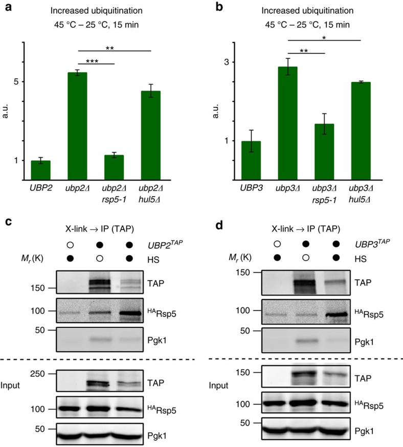
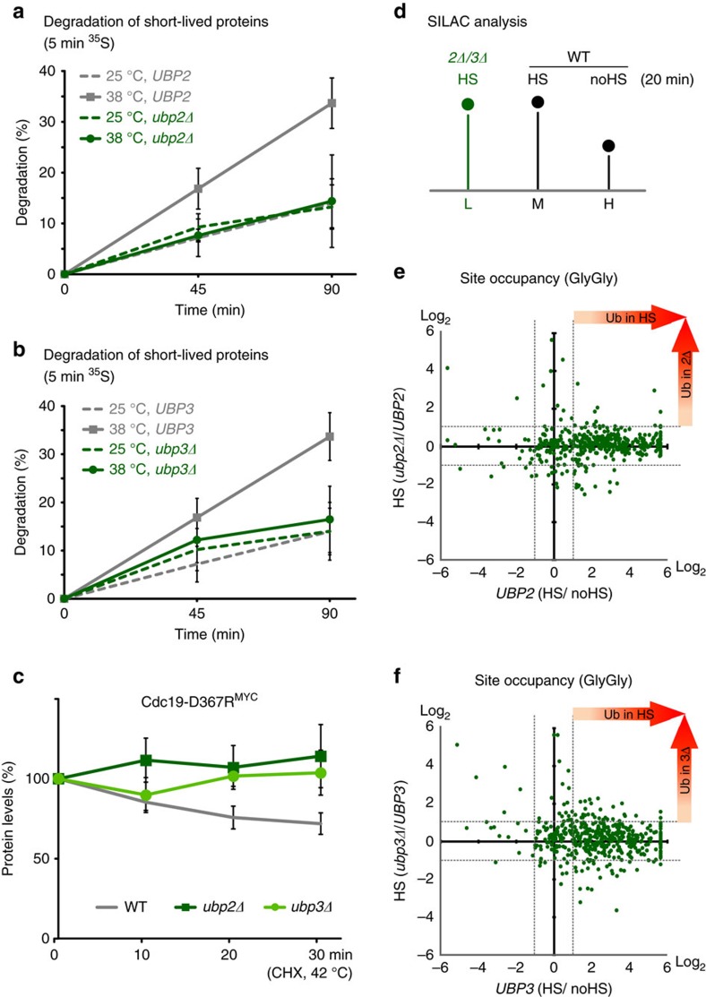
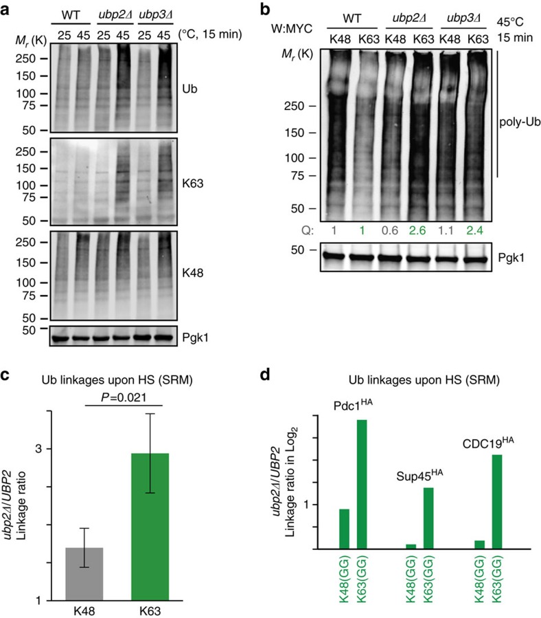
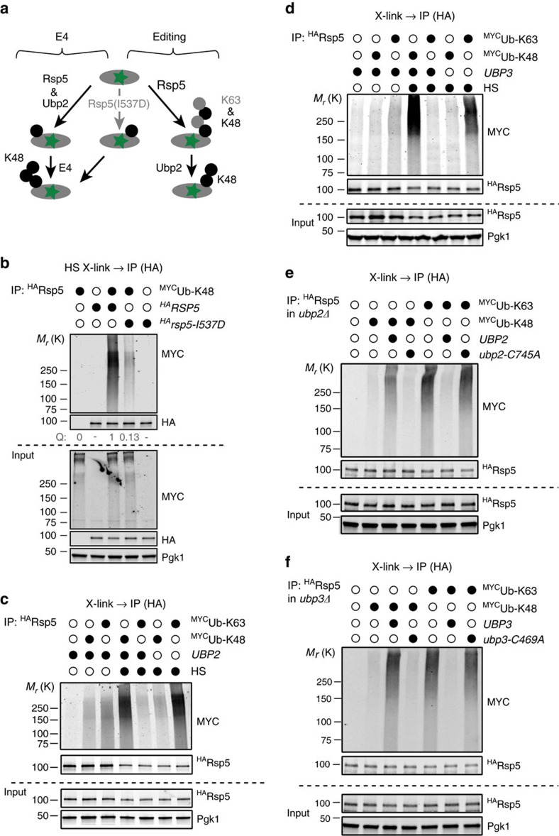
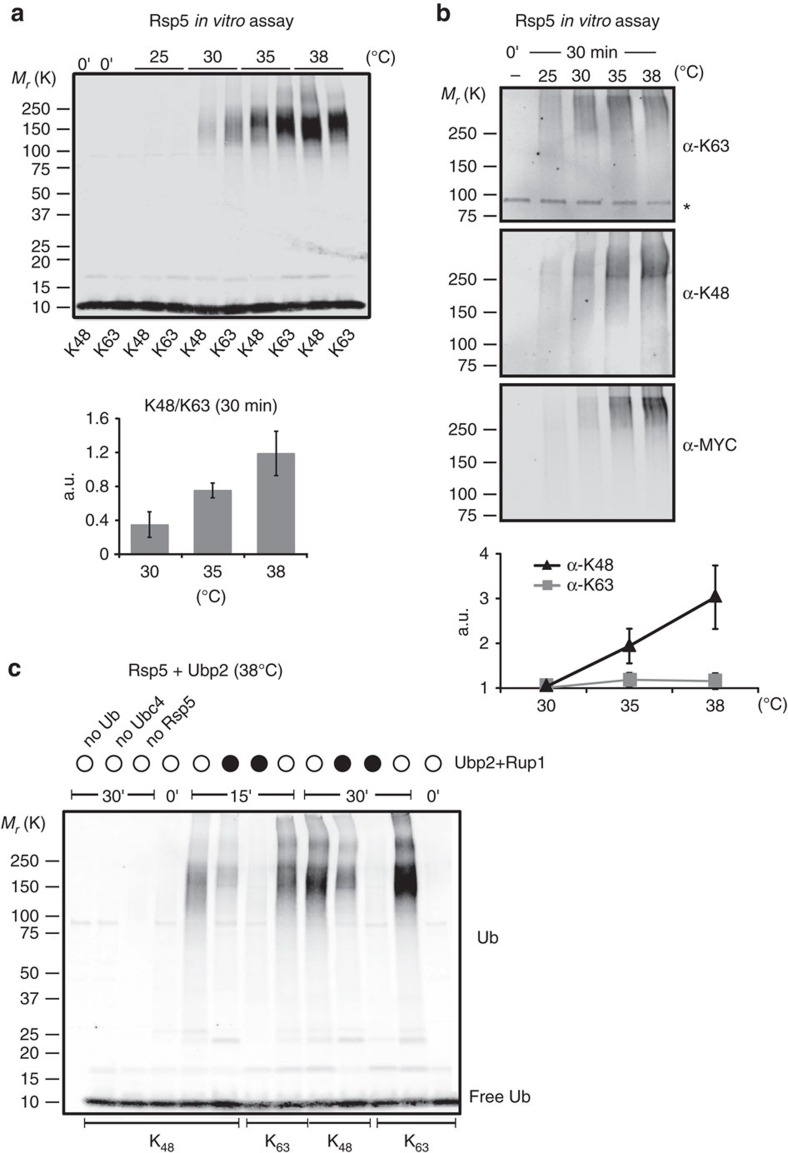
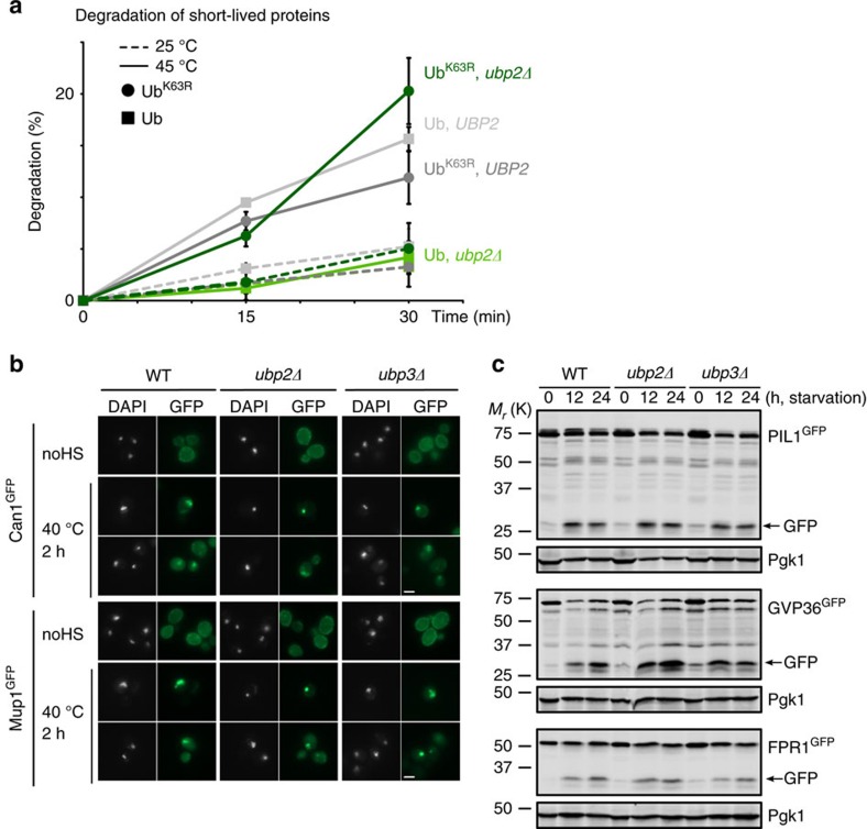
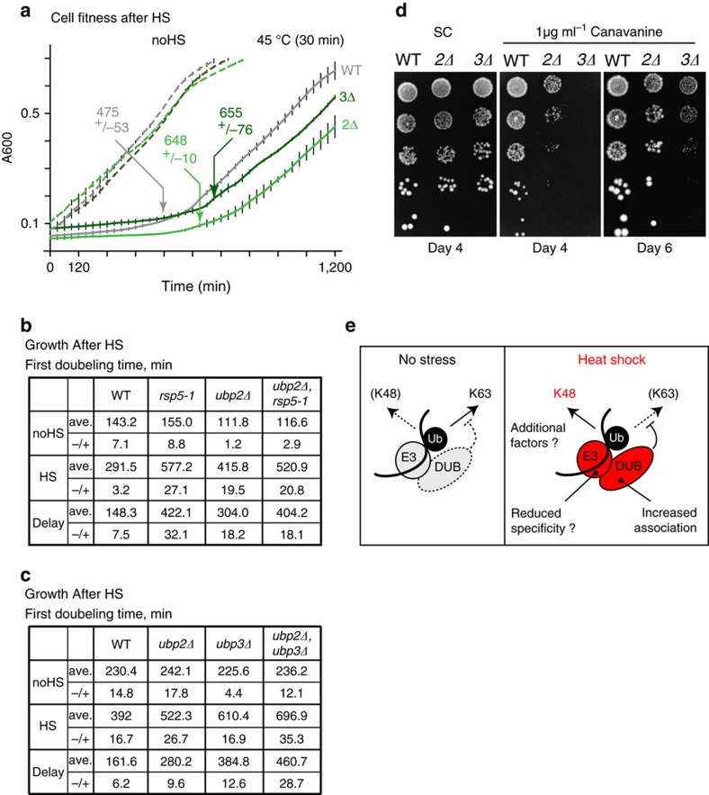
Elimination of misfolded proteins is crucial for proteostasis and to prevent proteinopathies. Nedd4/Rsp5 emerged as a major E3-ligase involved in multiple quality control pathways that target misfolded plasma membrane proteins, aggregated polypeptides and cytosolic heat-induced misfolded proteins for degradation. It remained unclear how in one case cytosolic heat-induced Rsp5 substrates are destined for proteasomal degradation, whereas other Rsp5 quality control substrates are otherwise directed to lysosomal degradation. Here we find that Ubp2 and Ubp3 deubiquitinases are required for the proteasomal degradation of cytosolic misfolded proteins targeted by Rsp5 after heat-shock (HS). The two deubiquitinases associate more with Rsp5 upon heat-stress to prevent the assembly of K63-linked ubiquitin on Rsp5 heat-induced substrates. This activity was required to promote the K48-mediated proteasomal degradation of Rsp5 HS-induced substrates. Our results indicate that ubiquitin chain editing is key to the cytosolic protein quality control under stress conditions.


Figures

Figure 1
Figure 1. Ubp2 and 3 control ubiquitination levels upon HS.
(a) Ubiquitination levels were quantified by dot-blots and normalized to Pgk1 levels in WT and the indicated single-deletion strains. Cells were maintained at 25 °C or incubated at 45 °C for 5 min. (b,c) Normalized ubiquitination levels were quantified by dot-blots (shown in Supplementary Fig. 1A) as in A and compared with a two-tail student t-test (**P<0.01). (d,e) Differences of normalized ubiquitination levels between unstressed cells (25 °C) and HS cells (45 °C for 5 min) quantified by dot-blots in the designated strains with the indicated plasmids (‘-' denotes control empty plasmid). Increased ubiquitination levels were compared using a two-tail student t-test (**P<0.01). (f) Differences of normalized ubiquitination levels before and after HS (45 °C for 5 min) quantified by dot-blots in the indicated cells. All experiments in bf were done with three biological replicates and averaged values are shown with s.d. Results are shown as arbitrary units (a.u.) and each value is relative to the reference sample.
Figure 2
Figure 2. Ubp2 and 3 mainly deubiquitinate Rsp5 substrates and have increased interactions with Rsp5 after HS.
(a,b) Increased ubiquitination levels after a 15 min HS at 45 °C in the indicated cells quantified by dot-blot and compared with unstressed cells (25 °C). Experiments were done with three biological replicates and averaged values are shown with s.d. In each experiment, increased ubiquitination levels were compared, as shown, to the deletion or WT strains using a two-tail student t-test (***P<0.001; **P<0.01; *P<0.05). a.u., arbitrary units. (c,d) Cells expressed 3xHA-tagged Rsp5 from a plasmid and endogenously C-terminally TAP-tagged UBP2 or UBP3, when indicated (black circles). Before lysis, cells that remained at 25 °C (unfilled circles) or heat shocked (40 °C, 20 min; black circles) were crosslinked for the remaining 10 min with 1% formaldehyde. Western blots of the indicated proteins are shown.
Figure 3
Figure 3. Ubp2 and 3 share substrate pools and are required for the degradation of misfolded proteins upon HS.
(a,b) Degradation of 35S pulsed-labelled proteins (5 min) in WT (grey) and indicated deletion strains (green) cells at 25 °C (dotted lines) or 38 °C (straight lines). The portion of proteins degraded at the indicated times was reported from three independent experiments with s.d. (c) Turnover of plasmid-expressed Cdc19-D367R13MYC in designated cells at 42 °C was assessed by western blots after the addition of 100 μg ml−1 cycloheximide. The Pgk1-normalized protein levels were averaged from three independent experiments and shown with s.d. (d) Schematic representation of the triple-SILAC experiment to quantify conjugated substrate peptides. L, M, and H denote the three different SILAC-labels (light, medium and heavy). (e,f) Scatter plots of the log2 ratios of quantified conjugated peptides. In the x axis, ratios of M versus H are shown to display sites affected by heat-stress in WT cells. In the y axis, ratios of L versus M are reported to display sites affected by the absence of UBP2 (e) or UBP3 (f) upon HS.
Figure 4
Figure 4. Ubp2 and 3 suppress the buildup of K63-linked chains after HS in vivo.
(a) Levels of K48- and K63-linked ubiquitin in designated cells were assessed using the chain-specific antibodies in cells grown at 25 °C and HS at 45 °C for 15 min. (b) Indicated cells that expressed MYCubiquitin-K48 only (K48) or MYCubiquitin-K63 only (K63) were incubated at 45 °C for 15 min. Quantified values (Q) of anti-Myc signal (>75 kDa) normalized to Pgk1 are shown relative to WT cells. (c) Levels of K48- and K63-linked ubiquitin in ubp2Δ and WT cells that expressed H8-ubiquitin under the GPD1 promoter were quantified by SRM after Ni2+ chromatography and SDS–PAGE separation. Cells were heat shocked at 45 °C for 20 min and ratios of levels between ubp2Δ and WT were measured in three independent experiments (with s.d.) using the indicated spiked-in labelled diGly peptides and compared with a two-tail student t-test. (d) The levels of K48- and K63-linked ubiquitin conjugated to the indicated Rsp5 substrates were quantified by SRM. After HS (45 °C for 20 min) endogenously C-terminally 3 × HA-tagged substrates were purified using the anti-HA antibody and separated by SDS–PAGE before in-gel trypsin digest of the high molecular weight species. Ratios of the indicated ubiquitin linkages between ubp2Δ and WT cells are shown.
Figure 5
Figure 5. Rsp5 conjugates K48-linked ubiquitin chains to its heat-induced substrates in vivo in an Ubp2 and 3-dependent manner.
(a) Schematic representation of two possible models to explain the role of Rsp5 in the assembly of K48 chains after HS. The I537D mutant of Rsp5 that impairs mostly poly- but not monoubiquitination should only strongly affect the editing pathway. (b) Cells that expressed 3HARsp5 or 3HARsp5-I537D together with empty or MYCubiquitin-K48 only constructs were heat shocked for 20 min (40 °C) and crosslinked with 1% formaldehyde for the remaining 10 min before lysis with SDS and the anti-HA IP. Western blot analysis of both IP and input samples are shown. (c,d) WT and ubp2Δ (c) or ubp3Δ (d) cells that expressed 3HARsp5 together with empty, MYCubiquitin-K48 only or MYCubiquitin-K63 only constructs were heat shocked (40 °C, 20 min) or not, and crosslinked with 1% formaldehyde before lysis with SDS and IP with anti-HA antibody-conjugated magnetic beads. Western blot analysis of both IP and Input samples are shown. (e,f) As in b,c, the ubp2Δ (e) or ubp3Δ (f) cells carried out either an empty control plasmid or a plasmid that expressed the WT or catalytically inactive deubiquitinases.
Figure 6
Figure 6. Rsp5 specificity is altered at higher temperatures and promotes K48 chains assembly in the presence of Ubp2 in vitro.
(a) In vitro Rsp5 auto-ubiquitination assay for 30 min at the indicated temperatures with His6-tagged K48-only (K48) or K63-only (K63) ubiquitin variants, followed by anti-ubiquitin western blots. Histograms (right) show quantification of K48 versus K63 levels (>100 kDa) from three independent experiments (with s.d.). Levels at 25 °C were too close to the background for quantification. (b) In vitro Rsp5 auto-ubiquitination assay at the indicated temperatures for 30 min with the Myc-tagged ubiquitin followed by western blots with anti- K63 chains, K48 chains and Myc antibodies. Asterisk denotes an unspecific band. Relative levels were quantified in three independent experiments (with s.d.). (c) In vitro Rsp5 auto-ubiquitination assay at 38 °C incubated for 15 or 30 min followed by western blot with anti-ubiquitin antibody. Unless indicated otherwise, the reactions were done in the presence of E1, Ubc4, Rsp5, His6-ubiquitin and an equimolar amount of Ubp2 and Rup1 (black circles).
Figure 7
Figure 7. Removal of K63 chains by Ubp2 and 3 mainly promotes proteasome degradation after HS.
(a) Degradation of 35S pulsed-labelled proteins in WT (grey) and ubp2Δ (green) cells at 25 °C (dotted lines) or 45 °C (straight lines) that expressed solely ubiquitin (light, square) or ubiquitin-K63R (dark, round). The portion of proteins degraded at the indicated times was measured and averaged for short-lived proteins in three independent experiments (with s.d.). (b) Representative fluorescent microscopy images of WT, ubp2Δ and ubp3Δ cells that expressed Can1GFP or Mup1GFP from their endogenous promoters and that were incubated at 25 or 40 °C for 2 h. Both the GFP and Hoechst (DNA staining) channels are shown. Scale bar, 2 μm. (c) Cells that expressed the indicated aggregation-prone proteins C-terminally tagged with GFP (from endogenous locus) were starved at 30 °C for the indicated times before western blots.
Figure 8
Figure 8. Absence of UBP2 or UBP3 reduces cell fitness after HS.
(a) The cell fitness of WT (grey), ubp2Δ (light green) and ubp3Δ (green) cells was measured in three biological replicates (horizontal bars represent s.d.). Cells were grown at 25 °C after no HS (dotted lines) or a HS at 45 °C for 30 min (straight lines). Time (min) required to complete the first doubling after HS is indicated for each strain with s.d. (b,c) Tables showing times (min) required to complete the first doubling with or without (45 °C, 30 min) treatment for the indicated cells. The experiment was done with three biological replicates (±s.d.). (d) 1/5 dilutions of WT, ubp2Δ and ubp3Δ cells grown on canavanine (1 μg ml−1) and control (SC) plates at 30 °C. (e) Proposed model in which Rsp5 assembles K63-linked chains in unstressed conditions, while it builds up K48-linked chains upon HS to signal for proteasomal degradation.

Similar articles

Cited by

References

    1. Ciechanover A. & Kwon Y. T. Degradation of misfolded proteins in neurodegenerative diseases: therapeutic targets and strategies. Exp. Mol. Med. 47, e147 (2015). - PMC - PubMed
    1. Comyn S. A., Chan G. T. & Mayor T. False start: cotranslational protein ubiquitination and cytosolic protein quality control. J. Proteomics 100, 92–101 (2014). - PubMed
    1. Powers E. T. & Balch W. E. Diversity in the origins of proteostasis networks-a driver for protein function in evolution. Nat. Rev. Mol. Cell Biol. 14, 237–248 (2013). - PMC - PubMed
    1. Valastyan J. S. & Lindquist S. Mechanisms of protein-folding diseases at a glance. Dis. Model. Mech. 7, 9–14 (2014). - PMC - PubMed
    1. Kleiger G. & Mayor T. Perilous journey: a tour of the ubiquitin-proteasome system. Trends Cell Biol. 24, 352–359 (2014). - PMC - PubMed

Publication types

MeSH terms

Grants and funding