. 2016 Oct 4:7:13019.
doi: 10.1038/ncomms13019.

Anti-tubulin drugs conjugated to anti-ErbB antibodies selectively radiosensitize

Affiliations

Anti-tubulin drugs conjugated to anti-ErbB antibodies selectively radiosensitize

Stephen R Adams et al. Nat Commun. .

Abstract

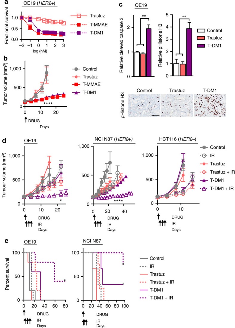
Tumour resistance to radiotherapy remains a barrier to improving cancer patient outcomes. To overcome radioresistance, certain drugs have been found to sensitize cells to ionizing radiation (IR). In theory, more potent radiosensitizing drugs should increase tumour kill and improve patient outcomes. In practice, clinical utility of potent radiosensitizing drugs is curtailed by off-target side effects. Here we report potent anti-tubulin drugs conjugated to anti-ErbB antibodies selectively radiosensitize to tumours based on surface receptor expression. While two classes of potent anti-tubulins, auristatins and maytansinoids, indiscriminately radiosensitize tumour cells, conjugating these potent anti-tubulins to anti-ErbB antibodies restrict their radiosensitizing capacity. Of translational significance, we report that a clinically used maytansinoid ADC, ado-trastuzumab emtansine (T-DM1), with IR prolongs tumour control in target expressing HER2+ tumours but not target negative tumours. In contrast to ErbB signal inhibition, our findings establish an alternative therapeutic paradigm for ErbB-based radiosensitization using antibodies to restrict radiosensitizer delivery.


Conflict of interest statement

University of California San Diego has filed patent applications based on the findings described in this manuscript (S.R.A., E.N.S., R.Y.T. and S.J.A.). The remaining authors declare no competing financial interests.

Figures

Figure 1
Figure 1. Anti-ErbB antibody MMAE conjugates bind in a receptor-dependent manner.
(a) CAL-27 (EGFR+) cells exposed to 2 nM of Cy5-labelled C-MMAE for 30 min then incubated in drug-free media. Cells were fixed at indicated times and imaged for Cy5 fluorescence (red). Nuclei stained with DAPI (blue). Scale bar, 10 μm. Representative images of three independent experiments. (b) A panel of EGFR (CAL27, A549, CALU3) and HER2 (CALU3, OE19, BT474) expressing cells from diverse tumour histologies were exposed to 2 nM Cy5-labelled C-MMAE or T-MMAE for 2 h and Cy5 fluorescence (red) imaged. Nuclei stained with DAPI (blue). Scale bar, 10 μm. Representative images of three independent experiments. (c) Cell surface binding of Cy5-labelled C-MMAE or T-MMAE. CAL-27 and OE19 cells incubated on ice with increasing concentrations of C-MMAE or T-MMAE. of Flow cytometry assessment of Cy5 signal. Representative data of two independent experiments. (d) Phase contrast microscopy of CAL-27 cells treated with 2 nM MMAE, cetuximab, or C-MMAE overnight. Representative images of three independent experiments. Scale bar, 50 μm. (e) Cell cycle profile of OE19 cells treated with MMAE, ErbB antibodies (cetuximab or trastuzumab) or ADC (C-MMAE or T-MMAE) overnight, stained with propidium iodide and analysed by flow cytometry. Data representative of two independent experiments.
Figure 2
Figure 2. Antibody conjugated MMAE selectively radiosensitizes tumour cells.
(a) CAL-27 (EGRF+) and OE19 (HER2+) cells were exposed to a dose range of cisplatin, paclitaxel or MMAE for 72–96 h. Cell viability normalized to vehicle treated cells and plotted as mean fractional survival±s.d. of three replicates. Data representative of three independent experiments. (b) CAL-27 and OE19 tumour cells were exposed to dose range of MMAE, ErbB antibody (cetuximab or trastuzumab) or ADC (C-MMAE or T-MMAE) for 72 h. Cell viability plotted as mean fractional survival±s.d. of three replicates. Data representative of three independent experiments. (c) Clonogenic cell survival (SF2) of CAL-27 cells treated with MMAE, cetuximab or C-MMAE overnight followed by 0 or 2 Gy. Cell viability normalized to non-irradiated cells for each drug condition and plotted as mean fractional survival±s.d. of six replicates. Data representative of two independent experiments. (d) CAL-27 and LN229 cells treated with cetuximab or C-MMAE overnight, irradiated with 6 Gy, and comet tail length measured by neutral comet assay. Data normalized to vehicle treated, non-irradiated cells and plotted as mean relative comet tail length±s.e.m. of >50 cells per group. Data representative of two independent experiments. (e) CAL-27 and LN229 cells exposed to 2 nM Cy5-labelled C-MMAE for 2 h and Cy5 fluorescence (red) imaged. Nuclei stained with DAPI (blue). Scale bar, 10 μm. (f) CALU3 (EGFR+, HER2+) and OE19 cells treated with free MMAE, ErbB antibodies (cetuximab or trastuzumab) or ADC (C-MMAE or T-MMAE) overnight, irradiated with 6 Gy, and comet tail length measured using neutral comet assay. Comet tail length normalized to vehicle treated, non-irradiated cells and plotted as mean relative comet tail length±s.e.m. of >50 cells per group. Data representative of two independent experiments. All statistical significances were calculated using one-way ANOVA with Tukey's multiple comparisons test. *P<0.05, **P<0.01, ***P<0.001. ****P<0.0001.
Figure 3
Figure 3. Clinical grade anti-tubulin ADC T-DM1 radiosensitizes HER2+ tumours.
(a) Clonogenic cell survival (SF2) of cells treated with 5 nM mertansine overnight followed by 2 Gy. Cell viability normalized to non-irradiated cells for each drug condition and plotted as mean fractional survival±s.d. of five replicates. Data representative of two independent experiments. (b) IC50 of mertansine and T-DM1 in cells based on HER2 expression. Cells treated with a dose range of drug for 72–96 h and viability measured, IC50 calculated and plotted as mean±s.d. of three independent experiments. (c) Cytotoxicity of clinically used radiosensitizers. OE19 cells were exposed to a dose range of drugs, viability normalized to vehicle treated cells and plotted as mean fractional survival±s.d. of three replicates. Data representative of three independent experiments (d) Cell cycle profile of OE19 and HCT116 cells treated with T-DM1 overnight, stained with propidium iodide and analysed by flow cytometry. Data representative of two independent experiments. (e) Neutral comet assay of HER2+ and HER2− cells treated with 20 nM mertansine, trastuzumab or T-DM1 overnight and irradiated with 2 Gy. Comet tail length was normalized to vehicle treated, non-irradiated cells and plotted as relative comet tail length±s.e.m. of >50 cells per group. Data representative of two independent experiments. (f) Representative images of comet tails and quantification of relative comet tail length of OE19 cells treated with 0–5 nM of T-DM1 and irradiated. Comet tail length was normalized to vehicle treated, non-irradiated cells and plotted as relative comet tail length±s.e.m. of >50 cells per group. Scale bar, 50 μm. All statistical significances were calculated using one-way ANOVA with Tukey's multiple comparisons test. *P<0.05, **P<0.01, ****P<0.0001.
Figure 4
Figure 4. Antibody conjugated MMAE selectively targets tumours and increases efficiency of tumour regression in combination with IR.
(a) OE19 tumour xenografts grown in the flank and bilateral thighs (location indicated by three red arrows) of mice and 0.5 nmol of Cy5-labelled C-MMAE or T-MMAE (∼4 and ∼3.9 mg kg−1, respectively) intravenously injected. Tumours imaged 48 h later for Cy5 fluorescence (upper left). Yellow arrowheads indicate gut auto-fluorescence. Tumour xenografts were harvested and stained for pHistone H3 (bottom left). From T-MMAE injected mice, tumours were H&E stained and imaged for Cy5 fluorescence (right). Scale bar, 50 μm. Representative imaging of two mice per group. (b) Mice were injected with Cy5-labelled T-MMAE, blood collected, Cy5 fluorescence measured and plotted as mean±s.d. of four mice. (c) EGFR+ tumour xenografts grown in the bilateral thighs of athymic nude mice (left), 0.5 nmoles of Cy5-labelled C-MMAE (∼4 mg kg−1) injected, 24 h later right tumour bearing thigh irradiated with 3 Gy and mice imaged 24 h post IR for Cy5 fluorescence. Non-irradiated, mice bearing unilateral CAL-27 tumour xenografts (right), 0.5 nmol of Cy5-labelled C-MMAE (∼4 mg kg−1) injected and mice imaged at 48 and 72 h for Cy5 fluorescence. Representative imaging of three mice per group. (d) EGFR+ tumour-bearing mice were injected with cetuximab or C-MMAE, harvested 24 h later and stained for pS10 histone H3. Scale bar, 50 μm. Representative imaging of three mice per group. (e) CAL-27 tumour-bearing mice were injected with 2 nmol of C-MMAE or T-MMAE (∼15.9 and ∼15.4 mg kg−1, respectively) and harvested. Tumour drug concentrations quantitated LC-MS/MS and plotted as mean±s.e.m. of four tumours. Statistical significance was calculated using one-way ANOVA with Tukey's multiple comparisons test. (f,g) Mice bearing CAL-27 tumours injected on day 0 with indicated drugs on day 0 and 3 Gy given on days 1, 2. Tumours were measured and plotted as mean tumour volume±s.e.m. of ≥8 tumours per group. Statistical significances were calculated using two-way ANOVA with Tukey's multiple comparisons test. **P<0.01, ****P<0.0001.
Figure 5
Figure 5. T-DM1 combined with IR results in long-term HER2+ tumour control.
(a) OE19 cells were exposed to trastuzumab, T-MMAE or T-DM1 for 96 h. Cell viability normalized to vehicle-treated cells and plotted as mean fractional survival±s.d. of three replicates. Data representative of three independent experiments. (b) Mice bearing OE19 tumour xenografts were i.v. injected on day 0 with 1 nmol trastuzumab, T-DM1 or T-MMAE (∼7.3, ∼7.4 and ∼7.7 mg kg−1, respectively). Tumours were measured twice a week and plotted as mean tumour volume±s.e.m. of 10 tumours per group. Statistical significances were calculated using two-way ANOVA with Tukey's multiple comparisons test. (c) Tumour from OE19 tumours harvested 48 h after trastuzumab or T-MMAE and stained for cleaved caspase 3 or p-Histone H3 by IHC. Data were normalized to untreated, control tumours and plotted as mean±s.e.m. of ≥3 replicates. Statistical significance was calculated using one-way ANOVA with Tukey's multiple comparisons test. Representative images of pHistone H3 shown. Scale bar, 75 μm. (d) Mice bearing OE19 or NCI N87 (HER2+) or HCT116 (HER2−) tumour xenografts were i.v. injected on day 0 with 0.25 nmol trastuzumab or T-DM1 (∼1.8 and ∼1.9 mg kg−1, respectively). IR-treated mice were given 2.5 Gy on days 1, 2 and 3. Tumours were measured twice a week and plotted as mean tumour volume±s.e.m. of ≥4 tumours per group. Statistical significances were calculated using two-way ANOVA with Tukey's multiple comparisons test. (e) Survival curve of HER2+ treated tumour xenografts of ≥2 mice per group . Statistical significances were calculated using log-rank (Mantel–Cox) test. *P<0.05, **P<0.01, ****P<0.0001.

References

    1. Marur S. & Forastiere A. A. Challenges of integrating chemotherapy and targeted therapy with radiation in locally advanced head and neck squamous cell cancer. Curr. Opin. Oncol. 222, 206–211 (2010). - PubMed
    1. Laine A. M., Westover K. D. & Choy H. Radiation therapy as a backbone of treatment of locally advanced non-small cell lung cancer. Semin. Oncol. 41, 57–68 (2014). - PubMed
    1. Cohen D. J. & Leichman L. Controversies in the treatment of local and locally advanced gastric and esophageal cancers. J. Clin. Oncol. 33, 1754–1759 (2015). - PubMed
    1. Herskovic A. et al.. Combined chemotherapy and radiotherapy compared with radiotherapy alone in patients with cancer of the esophagus. N. Engl. J. Med. 326, 1593–1598 (1992). - PubMed
    1. Epidermoid anal cancer: results from the UKCCCR randomised trial of radiotherapy alone versus radiotherapy, 5-fluorouracil, and mitomycin. UKCCCR Anal Cancer Trial Working Party. UK Co-ordinating Committee on Cancer Research. Lancet 348, 1049–1054 (1996). - PubMed

Publication types

MeSH terms