. 2016 Oct 4:6:34581.
doi: 10.1038/srep34581.

Apocynin and Nox2 regulate NF-κB by modifying thioredoxin-1 redox-state

Affiliations

Apocynin and Nox2 regulate NF-κB by modifying thioredoxin-1 redox-state

Silvia Cellone Trevelin et al. Sci Rep. .

Abstract

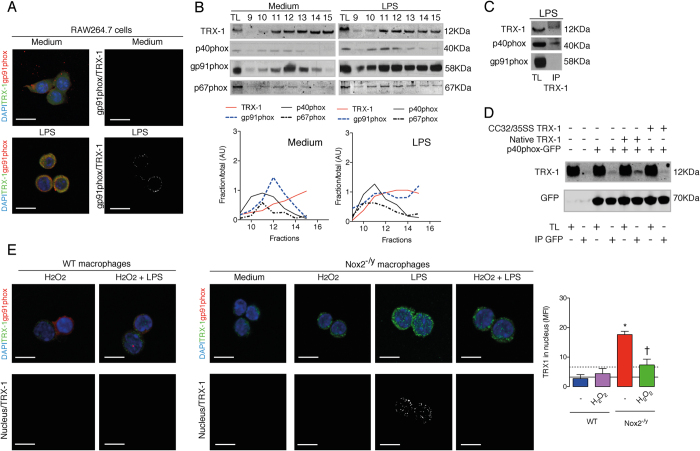
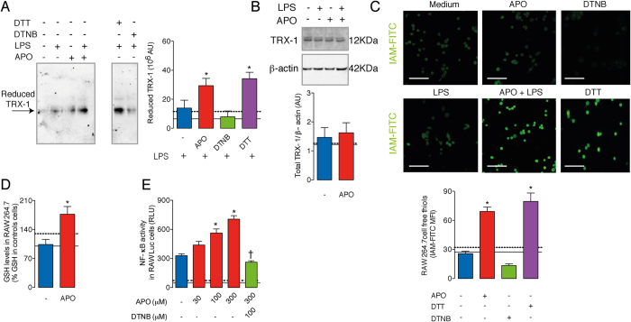
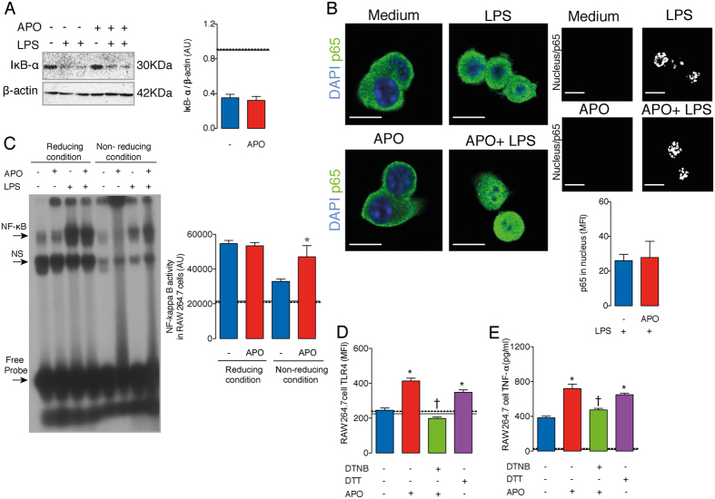
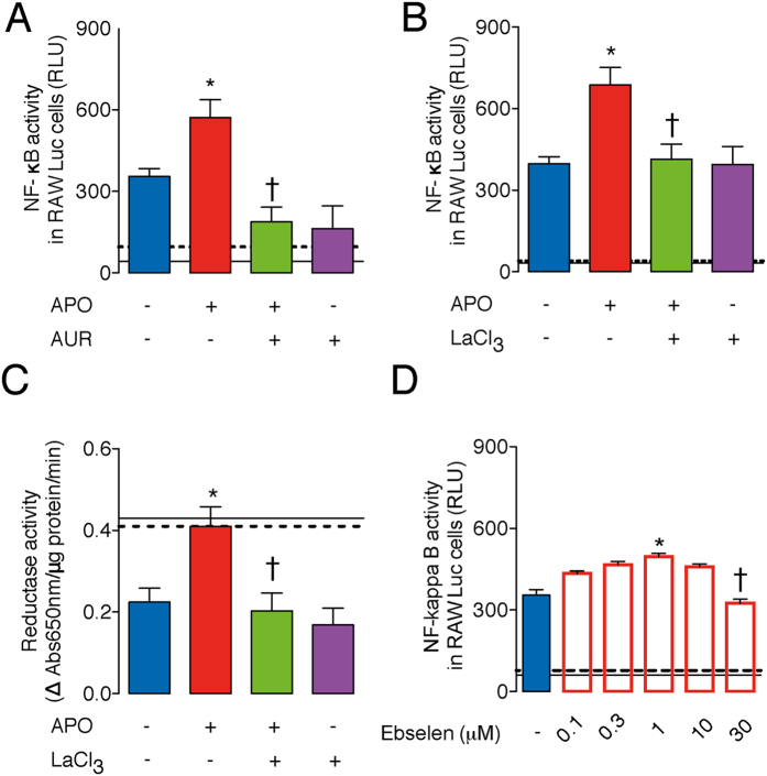
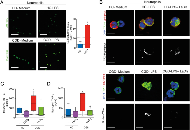
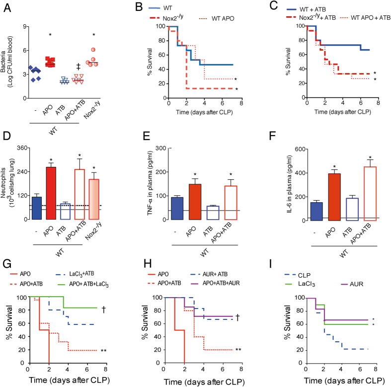
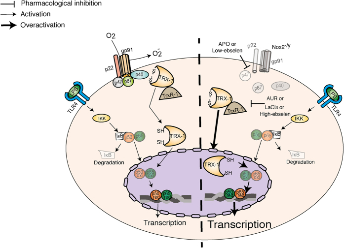
The reactive-oxygen-species-(ROS)-generating-enzyme Nox2 is essential for leukocyte anti-microbial activity. However its role in cellular redox homeostasis and, consequently, in modulating intracellular signaling pathways remains unclear. Herein, we show Nox2 activation favors thioredoxin-1 (TRX-1)/p40phox interaction, which leads to exclusion of TRX-1 from the nucleus. In contrast, the genetic deficiency of Nox2 or its pharmacological inhibition with apocynin (APO) results in reductive stress after lipopolysaccharide-(LPS)-cell stimulation, which causes nuclear accumulation of TRX-1 and enhanced transcription of inflammatory mediators through nuclear-factor-(NF)-κB. The NF-κB overactivation is prevented by TRX-1 oxidation using inhibitors of thioredoxin reductase-1 (TrxR-1). The Nox2/TRX-1/NF-κB intracellular signaling pathway is involved in the pathophysiology of chronic granulomatous disease (CGD) and sepsis. In fact, TrxR-1 inhibition prevents nuclear accumulation of TRX-1 and LPS-stimulated hyperproduction of tumor-necrosis-factor-(TNF)-α by monocytes and neutrophils purified from blood of CGD patients, who have deficient Nox2 activity. TrxR-1 inhibitors, either lanthanum chloride (LaCl3) or auranofin (AUR), also increase survival rates of mice undergoing cecal-ligation-and-puncture-(CLP). Therefore, our results identify a hitherto unrecognized Nox2-mediated intracellular signaling pathway that contributes to hyperinflammation in CGD and in septic patients. Additionally, we suggest that TrxR-1 inhibitors could be potential drugs to treat patients with sepsis, particularly in those with CGD.


Figures

Figure 1
Figure 1. Nox2 activation facilitates thioredoxin-1 (TRX-1) and p40phox interaction and prevents nuclear accumulation of TRX-1.
(A) TRX-1 (green) and Nox2/gp91phox (red) in RAW264.7 cells stimulated with LPS (10 ng/ml, 30 minutes). Nuclear material was stained with DAPI (blue). Images were obtained by confocal microscopy (63X objective; 5X magnification) and represent three independent experiments realized in triplet. Colocalization is highligted in white/black image. Scale bars, 7.5 μm. (B) Sucrose gradient cell fractionation and densitometry. Immunoblots (IB) were performed for TRX-1, p40phox, gp91phox and p67phox in fractions 9 to 15. AU: arbitrary units. (C) Immunoprecipitation (IP) was performed with anti-TRX-1 antibody (Ab) in total cell lysates and IB for p40phox, gp91phox and TRX-1. (D) HEK293T cells were transfected with p40phox-GFP and either native TRX-1 or mutated CC32/35SS TRX-1. IP was performed with anti-GFP Ab and IB for GFP and TRX-1. Blots represent three independent experiments. TL: total cell lysate. (E) Macrophages harvested from WT and Nox2−/y mice were stimulated with LPS. Some cells were treated with hydrogen peroxide (H2O2, 10 μM). Scale bars, 7.5 μm. The results are expressed as the means of fluorescence intensity (MFI) ±standard error of the mean (SEM) obtained by analyzing 15 nucleus-TRX-1 colocalizations (white) per group. Black continuous and dashed lines respectively indicate the average values obtained by analysing WT and Nox2−/y cells in medium only. Full-lengh blots are showed in Supplementary Figs 10 and 11. *P < 0.05 as compared to WT cells incubated with LPS; P < 0.05 as compared to Nox2−/y cells incubated with LPS.
Figure 2
Figure 2. Inhibition of Nox2 by APO results in TRX-1 reduction and in a reductive stress after LPS stimuli.
RAW264.7 cells were incubated with APO (300 μM, one hour) and stimulated with LPS (10 ng/ml, 30 minutes). Some cells were incubated with 5,5′-dithiobis-2-nitrobenzoic acid (DTNB, 100 μM) or dithiothreitol (DTT, 300 μM). Immunoblotting was performed under native (A) or reducing condition (B). Densitometry of three independent experiments. The results are expressed as the means ± SEM. AU: arbitrary units. (C) Levels of free thiols were determined by using IAM-FITC (green). The results are expressed as the means of fluorescence intensity (MFI) ± SEM obtained by analyzing 15–20 cells/group. Scale bars, 50 μm. (D) Levels of reduced glutathione (GSH) in total cell lysates. (E) RAW264.7-Luc cells were incubated with APO and then stimulated with LPS (10 ng/ml, four hours). Some cells were incubated with DTNB 30 minutes before the addition of APO. RLU: relative lumen units. Black continuous and dashed lines respectively indicate the average values obtained by analysing cells incubated in medium or after APO-treatment only. The results are expressed as the means ± SEM (n = 6/group, samples incubated with LPS; n = 3/group, samples without LPS). Full-lengh blots of the (B) are showed in Supplementary Fig. 12A. *P < 0.05 as compared to cells incubated with LPS; P < 0.05 as compared to LPS-stimulated cells pre-incubated with APO.
Figure 3
Figure 3. Inhibition of Nox2 by APO increases LPS-mediated NF-κB binding to DNA.
RAW264.7 cells were incubated with APO (300 μM, one hour) and then stimulated with LPS (10 ng/ml, 30 minutes). (A) Degradation of IκB-α, 15 minutes after LPS. Densitometry of three independent experiments. AU: arbitrary units. (B) Translocation of p65 (green) to nucleus (blue), 30 minutes after LPS. Images were acquired by confocal microscopy (63X objective; 5X magnification). Scale bars, 7.5 μm. The results are expressed as the means of fluorescence intensity (MIF) ±SEM obtained by analysing 15 nucleus-TRX-1 colocalizations (white) per group. (C) Eletromobility shift assay (EMSA) under reducing (plus DTT, 1 mM) or non-reducing conditions. NS: non-specific. (D) TLR4 expression by flow cytometry, 12 hours after LPS. (E) TNF-α levels in culture supernatants (ELISA), 12 hours after LPS. The results are expressed as the means ± SEM (n = 6/group, samples incubated with LPS; n = 3/group, samples without LPS). Black continuous and dashed lines respectively indicate the average values obtained by analysing cells incubated in medium or after APO-treatment only. Full-lengh blots of the (A) are showed in Supplementary Fig. 12B. *P < 0.05 as compared to cells incubated with LPS; P < 0.05 as compared to LPS-stimulated cells pre- incubated with APO.
Figure 4
Figure 4. TrxR-1 inhibition prevents the LPS-mediated NF-κB over activation induced by APO.
RAW264.7-Luc cells were incubated with APO (300 μM, one hour) and then stimulated with LPS (10 ng/ml, four hours). (A,B) Cells incubated with the TrxR-1 inhibitors, auranofin (AUR, 1 μM- [A]) or lanthanum chloride (LaCl3, 1 μM- [B]), for 30 minutes before APO. RLU: relative lumen units. (C) Reductase activity of cell lysates 30 minutes after LPS. (D) Cells were incubated with ebselen one hour before LPS. The results are expressed as the means ± SEM (n = 6/group, samples incubated with LPS; n = 3/group, samples without LPS). Black continuous and dashed lines indicate the average values obtained by analysing cells incubated in medium or APO-treatment (or ebselen-treatment 30 μM in (D), respectively. *P < 0.05 as compared to cells incubated with LPS; P < 0.05 as compared to LPS-stimulated cells pre- incubated with APO.
Figure 5
Figure 5. TrxR-1 inhibition prevents aberrant TNF-α production by leukocytes from chronic granulomatous disease patients (CGD) stimulated with LPS.
(AC) Neutrophils from healthy controls (HC, n = 5) or CGD patients (n = 5) were stimulated with LPS (10 ng/ml). (A) Staining with IAM-FITC (green), 30 minutes after LPS. Images were obtained by confocal microscopy (63X objective). Scale bars, 50 μm. The results are expressed as the means fluorescence intensity (MIF) ± SEM obtained by analyzing 15 cells/subject. (B) Colocalization (white) of TRX-1 (green) and Nox2 (gp91phox, red) in HC cells and nuclear accumulation of TRX-1 (white) in cells of CGD cells, 30 minutes after LPS. Nuclear material was stained with DAPI (blue). Images were obtained by confocal microscopy (63X objective; 5X magnification). Scale bars, 7.5 μm. (C,D) TNF-α levels in cultures supernatant (ELISA), 12 hours after LPS. Some cells were treated with lanthanum chloride (LaCl3, 1 μM) 30 minutes before LPS. Box plots show median, interquartile range, sample minimum and maximum. Black continuous and dashed lines indicate the average values obtained by analysing, respectively, HC and CGD cells in medium only. *P < 0.05 as compared to HC cells stimulated with LPS; P < 0.05 as compared to CGD cells stimulated with LPS.
Figure 6
Figure 6. TrxR-1 inhibition enhances sepsis survival of mice treated with APO.
Wild type (WT) mice subcutaneously treated with APO (200 mg/kg, 30 minutes before surgery) and Nox2−/y mice were submitted to cecal ligation and puncture (CLP). Some mice were intraperitonously treated with antibiotic (ATB, ertapenem sodium, 30 mg/kg), six hours after surgery and 12 hourly thereafter. (A) Bacterial load in blood, 12 hours after CLP. The graph represents the individual logarithmic values of CFU around median (n = 5–7/group). (B,C) Survival rates (n = 15/group). (DF) Number of neutrophils trapped in lungs, levels of TNF-α and IL-6 in plasma, six hours after ATB treatment and 12 hours after CLP. The results are expressed as the means ± SEM (n = 5–7/group). Black continuous and dashed lines in bar graphs indicate the average values obtained by analysing WT and Nox2−/y sham-operated mice, respectively. (GI) Mice were treated with LaCl3 (10 mg/kg, subcutaneously), which was administered six hours after surgery and 12 hourly thereafter; or mice were treated with auranofin (AUR, 2 mg/kg, subcutaneously), which was administered six hours after surgery and 24 hourly thereafter. Survival rates (n = 7–10/group; n = 7, AUR and/or APO treated mice; n = 10, LaCL3 and or APO treated mice). *P < 0.05 as compared to WT-CLP mice; P < 0.05 as compared to APO-CLP mice under antibiotic therapy; P < 0.05 as compared to APO-CLP mice without ATB treatment; **P < 0.05 as compared to LaCl3 -CLP or AUR -CLP mice, both under antibiotic therapy.
Figure 7
Figure 7. Nox2 regulates NF-κB activation by modifying TRX-1 redox-state.
Schematic representation. Left panel: Nox2 assembly favors TRX-1 interaction with p40phox. Oxidized TRX-1 is excluded from the nucleus by p40phox until its reduction by thioredoxin reductase-1 (TrxR-1). The TRX-1 reduction allows its nuclear localization, where it facilitates NF-κB binding to DNA. Right panel: the genetic deficiency (Nox2−/y) or pharmacological inhibition of Nox2 (with apocynin, APO; or low-concentration-ebselen) results in reduction of TRX-1 and its nuclear accumulation. In the nucleus, reduced TRX-1 potentiates NF-κB binding to DNA, and consequently, enhances the transcription of inflammatory mediators. TrxR-1 inhibitors, such as lanthanum chloride (LaCl3), auranofin (AUR) or high-concentration-ebselen prevent overactivation of NF-κB.

References

    1. Dinauer M. C. & Orkin S. H. Chronic granulomatous disease. Annu Rev Med. 43, 117–124 (1992). - PubMed
    1. Pollock J. D. et al.. Mouse model of X-linked chronic granulomatous disease, an inherited defect in phagocyte superoxide production. Nat Genet. 9, 202–209 (1995). - PubMed
    1. Deffert C. et al.. Hyperinflammation of chronic granulomatous disease is abolished by Nox2 reconstitution in macrophages and dendritic cells. J Pathol. 228, 341–350 (2012). - PubMed
    1. Whitmore L. C. et al.. NOX2 protects against prolonged inflammation, lung injury, and mortality following systemic insults. J Innate Immun. 5, 565–580 (2013). - PMC - PubMed
    1. Han W. et al.. NADPH oxidase limits lipopolysaccharide-induced lung inflammation and injury in mice through reduction-oxidation regulation of NF-kappaB activity. J Immunol. 190, 4786–4794 (2013). - PMC - PubMed

Publication types

MeSH terms

LinkOut - more resources