Specific clinical manifestations of Nocardia: A case report and literature review
- PMID: 27698688
- PMCID: PMC5038476
- DOI: 10.3892/etm.2016.3571
Specific clinical manifestations of Nocardia: A case report and literature review
Abstract
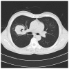
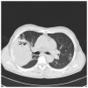
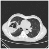
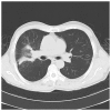
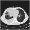
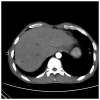
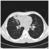
Nocardiosis is a rare bacterial infection of either the lungs (pulmonary) or body (systemic) that usually affects immunocompromised individuals. It is caused by Gram-positive, aerobic actinomycetes of the Nocardia genus. Multiple high-density sheet shadows in both lungs along with nodules or cavities are the most common presentations of nocardiosis, whereas a large pulmonary mass is considered to be rare. However, there is no specificity in the clinical manifestation of the disease. Therefore, isolation and identification of Nocardia strains is the only reliable diagnostic method. The present study describes the cases of two male patients of Asian descent with nocardiosis. Chest computed tomography scans showed a suspected tumor mass in both patients. Microscopic analysis and culturing of tissue samples obtained using a bronchoscope detected the presence of Nocardia wallacei. Neither patient showed signs of immunosuppression. The present study aimed to improve the understanding of lung nocardiosis and demonstrated that pulmonary nocardiosis should be suspected in the case of non-immunocompromised patients with a large mass in the lung. Furthermore, a review of the literature on infection with Nocardia was conducted.
Keywords: clinical manifestation; nocardia; opportunistic infection; pulmonary nocardiosis.
Figures



Similar articles
-
[Pulmonary nocardiosis in immunocompromised host: report of 2 cases and review of the literature].Zhonghua Jie He He Hu Xi Za Zhi. 2009 Aug;32(8):593-7. Zhonghua Jie He He Hu Xi Za Zhi. 2009. PMID: 19958678 Review. Chinese.
-
Endobronchial Enigma: A Clinically Rare Presentation of Nocardia beijingensis in an Immunocompetent Patient.Case Rep Pulmonol. 2015;2015:970548. doi: 10.1155/2015/970548. Epub 2015 Dec 24. Case Rep Pulmonol. 2015. PMID: 26819795 Free PMC article.
-
A complicated infection by cutaneous Nocardia wallacei and pulmonary Mycobacterium abscessus in a Chinese immunocompetent patient: a case report.Front Cell Infect Microbiol. 2023 Aug 16;13:1229298. doi: 10.3389/fcimb.2023.1229298. eCollection 2023. Front Cell Infect Microbiol. 2023. PMID: 37655298 Free PMC article.
-
A complicated case of an immunocompetent patient with disseminated nocardiosis.Infect Dis Rep. 2014 Apr 4;6(1):5327. doi: 10.4081/idr.2014.5327. eCollection 2014 Feb 18. Infect Dis Rep. 2014. PMID: 24757510 Free PMC article.
-
Lymphocutaneous type of nocardiosis caused by Nocardia brasiliensis: a case report and review of primary cutaneous nocardiosis caused by N. brasiliensis reported in Japan.J Dermatol. 2008 Jun;35(6):346-53. doi: 10.1111/j.1346-8138.2008.00482.x. J Dermatol. 2008. PMID: 18578712 Review.
Cited by
-
Genomic characterization of Nocardia seriolae strains isolated from diseased fish.Microbiologyopen. 2019 Mar;8(3):e00656. doi: 10.1002/mbo3.656. Epub 2018 Aug 16. Microbiologyopen. 2019. PMID: 30117297 Free PMC article.
-
Genotypic and phenotypic prevalence of Nocardia species in Iran: First systematic review and meta-analysis of data accumulated over years 1992-2021.PLoS One. 2021 Jul 22;16(7):e0254840. doi: 10.1371/journal.pone.0254840. eCollection 2021. PLoS One. 2021. PMID: 34292995 Free PMC article.
-
Ventilator-dependent pulmonary nocardiosis in a patient with chronic obstructive pulmonary disease.Lung India. 2018 Jan-Feb;35(1):58-61. doi: 10.4103/lungindia.lungindia_170_17. Lung India. 2018. PMID: 29319037 Free PMC article.
-
Pulmonary Infections Caused by Emerging Pathogenic Species of Nocardia.Case Rep Infect Dis. 2019 Oct 1;2019:5184386. doi: 10.1155/2019/5184386. eCollection 2019. Case Rep Infect Dis. 2019. PMID: 31662925 Free PMC article.
-
Clinical Nocardia species: Identification, clinical characteristics, and antimicrobial susceptibility in Shandong, China.Bosn J Basic Med Sci. 2020 Nov 2;20(4):531-538. doi: 10.17305/bjbms.2020.4764. Bosn J Basic Med Sci. 2020. PMID: 32415818 Free PMC article.
References
-
- Tuo MH, Tsai YH, Tseng HK, Wang WS, Liu CP, Lee CM. Clinical experiences of pulmonary and bloodstream nocardiosis in two tertiary care hospitals in northern Taiwan, 2000-2004. J Microbiol Immunol Infect. 2008;41:130–136. - PubMed
-
- Wu BQ, Zhang TT, Zhu JX, Liu H, Huang J, Zhang WX. Pulmonary nocardiosis in immunocompromised host: Report of 2 cases and review of the literature. Zhonghua Jie He He Hu Xi Za Zhi. 2009;32:593–597. (In Chinese) - PubMed
LinkOut - more resources
Full Text Sources
Other Literature Sources
