. 2016 Oct;12(4):2069-2077.
doi: 10.3892/etm.2016.3602. Epub 2016 Aug 22.

Diagnosing pyogenic, brucella and tuberculous spondylitis using histopathology and MRI: A retrospective study

Affiliations

Diagnosing pyogenic, brucella and tuberculous spondylitis using histopathology and MRI: A retrospective study

Tao Li et al. Exp Ther Med. 2016 Oct.

Abstract

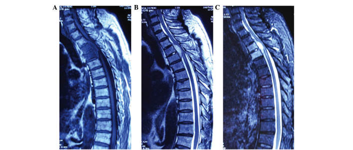
The present study examined the histopathological and magnetic resonance imaging (MRI) features of pyogenic, brucella and tuberculous spondylitis (PS, BS and TS, respectively). A total of 22 PS, 20 BS and 20 TS patients were included in the study. Histopathological examination was used to assess the lesion structure and composition, and the MRI observation identified the lesion location and signal features. The following histopathological and MRI features were identified significantly more in patients with PS than in patients with BS and TS: Predominant neutrophil infiltration, abnormal intervertebral disk signal, lesions on the ventral and lateral sides of the vertebral bodies, and thick and irregular abscess walls. The following histopathological and MRI features were identified significantly more in patients with BS than in patients with PS and TS: Predominant lymphocyte infiltration, new bone formation, epithelioid granuloma, lesions on the ventral sides of the vertebral bodies, no, or very mild, vertebral body deformation, no abnormal paraspinal soft tissue signal, no intraosseous or paraspinal abscesses, and thin and irregular abscess walls. The following histopathological and MRI features were identified significantly more in patients with TS than in patients with BS and PS: Sequestrum, Langerhans giant cells, caseous necrosis, lesions primarily in the thoracic region and on the lateral sides of the vertebral bodies, no obvious intervertebral disk damage, obvious vertebral body deformation, abnormal paraspinal soft tissue signal, intraosseous or paraspinal abscesses, and thin and smooth abscess walls. In conclusion, it can be suggested that these significant differences in histopathological and MRI features between the three different types of spondylitis may contribute towards the differential diagnosis of the diseases.

Keywords: brucella spondylitis; histopathology; magnetic resonance imaging; pyogenic spondylitis; tuberculous spondylitis.


Figures

Figure 1.
Figure 1.
Representative histopathological examination of bone tissue specimens from patients with pyogenic spondylitis (hematoxylin and eosin staining). (A) Clot and granulation tissues, without sequestrum or new bone formation (×100). (B) Predominant neutrophil infiltration (×400).
Figure 2.
Figure 2.
Representative histopathological examination of bone tissue specimens from patients with brucella spondylitis (hematoxylin and eosin staining). (A) Sequestrum, new bone formation and epithelioid granuloma, without Langerhans giant cells or caseous necrosis (×100). (B) Predominant lymphocyte infiltration (×400).
Figure 3.
Figure 3.
Representative histopathological examination of bone tissue specimens from patients with tuberculous spondylitis (hematoxylin and eosin staining). Sequestrum, epithelioid granuloma and caseous necrosis. at (A) ×100 and (B) ×400 magnification.
Figure 4.
Figure 4.
Magnetic resonance imaging examination of patients with pyogenic spondylitis in the lumbar region. (A) Hypointense signal on T1-weighted images, (B) hyperintense signal on T2-weighted images and (C) intermediate signal on enhanced T1 magnetic resonance in the lumbar region. A well-defined abnormal intervertebral disk signal and lesions on the ventral side of the vertebral body with unclear boundaries are present, without epidural abscesses.
Figure 5.
Figure 5.
Magnetic resonance imagine examination of patients with pyogenic spondylitis in the cervical region. (A) Hypointense signal on T1-weighted images, (B) intermediate signal on T2-weighted images and (C) hyperintense signal on enhanced T1 MR in the cervical region. A well-defined abnormal intervertebral disk signal, vertebral body deformation, lesions on the ventral side of the vertebral body with unclear boundary and epidural abscess are present.
Figure 6.
Figure 6.
Magnetic resonance imaging examination of patients with brucella spondylitis in the lumbar region. (A) Hypointense signal on T1-weighted images and (B) hyperintense signal on T2-weighted images are observed in the lumbar region. A normal intervertebral disk and limited lesions on the ventral side of the vertebral body with unclear boundaries are present, without epidural abscesses.
Figure 7.
Figure 7.
Magnetic resonance imaging examination of patients with brucella spondylitis in the thoracic region. (A) Hypointense signal on T1-weighted images, (B) intermediate-hyperintense signal on T2-weighted images and (C) hyperintense signal on T2-weighted images with fat suppression in the thoracic region. Abnormal intervertebral disks and epidural abscesses are present.
Figure 8.
Figure 8.
Magnetic resonance imaging examination of patients with tuberculous spondylitis in the thoracic region. (A) Hypointense signal on T1-weighted images and (B) intermediate-hyperintense signal on T2-weighted images of the thoracic region. A normal intervertebral disk, obvious vertebral body deformation, and abnormal paraspinal soft tissue with thin and smooth abscess walls are present.
Figure 9.
Figure 9.
Magnetic resonance imaging examination of patients with tuberculous spondylitis in the thoracic region. (A) Hypointense signal on T1-weighted images and (B) intermediate-hyperintense signal on T2-weighted images of the thoracic region. Abnormal intervertebral disks and normal paraspinal soft tissue, without obvious abscess, are present.

References

    1. Cheung WY, Luk KD. Pyogenic spondylitis. Int Orthop. 2012;36:397–404. doi: 10.1007/s00264-011-1384-6. - DOI - PMC - PubMed
    1. Sapico FL, Montgomerie JZ. Pyogenic vertebral osteomyelitis: Report of nine cases and review of the literature. Rev Infect Dis. 1979;1:754–776. doi: 10.1093/clinids/1.5.754. - DOI - PubMed
    1. Solera J, Lozano E, Martínez-Alfaro E, Espinosa A, Castillejos ML, Abad L. Brucellar spondylitis: Review of 35 cases and literature survey. Clin Infect Dis. 1999;29:1440–1449. doi: 10.1086/313524. - DOI - PubMed
    1. Colmenero JD, Cisneros JM, Orjuela DL, Pachón J, Garcia-Portales R, Rodriguez-Sampedro F, Juarez C. Clinical course and prognosis of Brucella spondylitis. Infection. 1992;20:38–42. doi: 10.1007/BF01704893. - DOI - PubMed
    1. Wang Dali, Zhang Shiyi. Advances in treatment of acute brucellosis. Zhong Guo Gan Ran Kong Zhi Za Zhi. 2001;16:94–95. (In Chinese)

LinkOut - more resources