. 2016 Oct;12(4):2671-2680.
doi: 10.3892/etm.2016.3641. Epub 2016 Aug 31.

Identification of the protective effects of traditional medicinal plants against SDS-induced Drosophila gut damage

Affiliations

Identification of the protective effects of traditional medicinal plants against SDS-induced Drosophila gut damage

Yang Zhou et al. Exp Ther Med. 2016 Oct.

Abstract

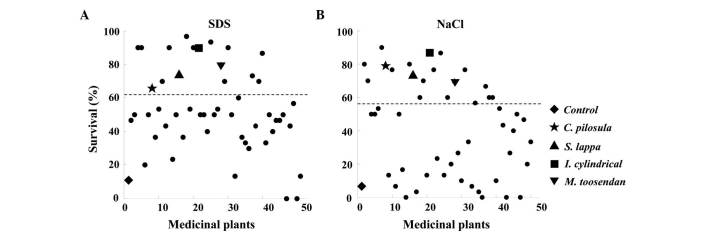
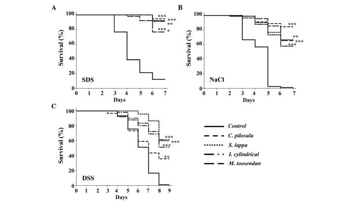
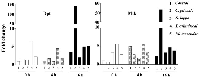
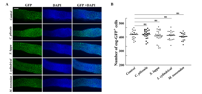
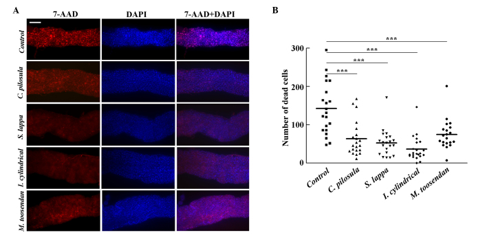
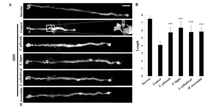
Traditional medicinal plants are widely used as immunomodulatory medicines that help improve health. A total of 50 different plants used for the treatment of toxicity were screened for their in vivo protective effects. Flies were fed a standard cornmeal-yeast medium (control group) or the standard medium containing medicinal plant extracts (experimental groups). Assessment of the survival rate was performed by feeding flies with toxic compounds. Gut epithelial cells were analyzed for cell proliferation and death by green fluorescent protein antibodies and 7-aminoactinomycin D staining under the microscope. The expression of antimicrobial peptides (AMPs) was evaluated by the quantitative polymerase chain reaction and the results revealed that after feeding the flies with toxic compounds, aqueous extracts from Codonopsis pilosula (Franch.) Nannf (C. pilosula), Saussurea lappa (Decne.) C.B.Clarke (S. lappa), Imperata cylindrica Beauv.var.major (Nees) C.E. Hubb. (I. cylindrical var. major) and Melia toosendan Sied. Et Zucc. (M.toosendan) increased the fly survival rate, reduced epithelial cell death and improved gut morphology. In addition, C. pilosula extracts induced the antimicrobial peptide levels (Dpt and Mtk) following treatment with sodium dodecyl sulfate (SDS). However, these extracts were not observed to increase SDS-induced cell proliferation in vivo. These results indicate that there are strong protective effects in extracts of C. pilosula, S. lappa, I. cylindrical var. major and M. toosendan on Drosophila intestinal cells among 50 medicinal plants.

Keywords: Drosophila melanogaster cell death; gut immunity; survival; traditional medicinal plant.


Figures

Figure 1.
Figure 1.
In vivo screening of the effects of anti-inflammatory reagents on the effect of 50 different traditional medicinal plant extracts. Survival rate on day 6 following treatment with (A) 0.6% SDS or (B) 0.4 M NaCl compared with the control group. The list of the 50 different traditional medicinal plants is shown in Tables III and IV. SDS, sodium dodecyl sulfate.
Figure 2.
Figure 2.
Survival rates of the control and experimental groups following treatment with SDS, NaCl or DSS. Adult flies cultured in standard medium (control) or supplemented with C. pilosula, S. lappa, I. cylindrical var. major and M. toosendan extracts were treated with 5% sucrose containing (A) 0.6% SDS, (B) 0.4 M NaCl or (C) 4% DSS at 25°C. The survival curves were derived from three independent experiments. **P<0.005 and ***P<0.001 vs. the control group. SDS, sodium dodecyl sulfate; DSS, dextran sulfate sodium.
Figure 3.
Figure 3.
RNA expression levels of antimicrobial peptides (AMPs) in the adult gut. Quantitative polymerase chain reaction analysis of AMPs in adult female guts isolated from flies that were treated with sodium dodecyl sulfate for 0, 4 or 16 h. Similar expression patterns were observed in two independent experiments. Dpt, Diptericin; Mtk, Metchnikowin.
Figure 4.
Figure 4.
Sodium dodecyl sulfate (SDS) treatment induces a change in the number of intestinal stem cells and enteroblasts in the Drosophila gut. (A) Expression of GFP under the control of the esg-Gal4 UAS-GFP specific reporter gene in intestinal stem cells (ISCs) and enteroblasts (EBs) was induced with 0.6% SDS for 16 h. esg-GFP, ISCs and EBs (green); DAPI, nucleus (blue); GFP + DAPI, green and blue. Scale bar, 100 µm. (B) Quantification of ISCs and EBs in the posterior midguts following SDS treatment; ≥20 guts were used per group. Error bars in the graph indicate the mean ± standard error of the mean. ns, no significant difference; GFP, green fluorescent protein; DAPI, 4′,6-diamidino-2-phenylindole.
Figure 5.
Figure 5.
Protective effects of medicinal plant extracts against epithelial cell death in the Drosophila gut following treatment with sodium dodecyl sulfate (SDS). (A) The guts of control and experimental flies were stained with 7-AAD following treatment with 0.6% SDS for 96 h. 7-AAD, dead cells (red); DAPI, nucleus (blue); 7-AAD + DAPI, red and blue. Scale bar, 100 µm. (B) Quantification of dead cells in the anterior midgut following SDS treatment; ≥20 guts were used per group. Error bars in the graph indicate the mean ± standard error of the mean. ***P<0.001. SDS, sodium dodecyl sulfate; AAD, aminoactinomycin; DAPI, 4′,6-diamidino-2-phenylindole; 7-AAD, 7-aminoactinomycin D.
Figure 6.
Figure 6.
Protective effects of medicinal plant extracts against morphological changes in the Drosophila gut following treatment with SDS. (A) Nomarski images of the Drosophila gut following treatment with 5% sucrose (negative control group) or 0.6% SDS dissolved in 5% sucrose for 4 days. Scale bar, 500 µm. The melanotic mass is shown in the magnified image of the square box of the control group. (B) The relative lengths of guts in panel (A). Error bars represent the standard deviation. ***P<0.001 vs. the control group. SDS, sodium dodecyl sulfate.

References

    1. Gautam R, Jachak SM. Recent developments in anti-inflammatory natural products. Med Res Rev. 2009;29:767–820. doi: 10.1002/med.20156. - DOI - PubMed
    1. Lee KA, Lee WJ. Drosophila as a model for intestinal dysbiosis and chronic inflammatory diseases. Dev Comp Immunol. 2014;42:102–110. doi: 10.1016/j.dci.2013.05.005. - DOI - PubMed
    1. Chatterjee M, Ip YT. Pathogenic stimulation of intestinal stem cell response in Drosophila. J Cell Physiol. 2009;220:664–671. doi: 10.1002/jcp.21808. - DOI - PMC - PubMed
    1. Micchelli CA, Perrimon N. Evidence that stem cells reside in the adult Drosophila midgut epithelium. Nature. 2006;439:475–479. doi: 10.1038/nature04371. - DOI - PubMed
    1. Jiang H, Patel PH, Kohlmaier A, Grenley MO, McEwen DG, Edgar BA. Cytokine/Jak/Stat signaling mediates regeneration and homeostasis in the Drosophila midgut. Cell. 2009;137:1343–1355. doi: 10.1016/j.cell.2009.05.014. - DOI - PMC - PubMed

LinkOut - more resources