. 2016 Oct;12(4):2389-2394.
doi: 10.3892/ol.2016.4921. Epub 2016 Jul 29.

Entrance of the Tat protein of HIV-1 into human uterine cervical carcinoma cells causes upregulation of HPV-E6 expression and a decrease in p53 protein levels

Affiliations

Entrance of the Tat protein of HIV-1 into human uterine cervical carcinoma cells causes upregulation of HPV-E6 expression and a decrease in p53 protein levels

Giovanni Barillari et al. Oncol Lett. 2016 Oct.

Abstract

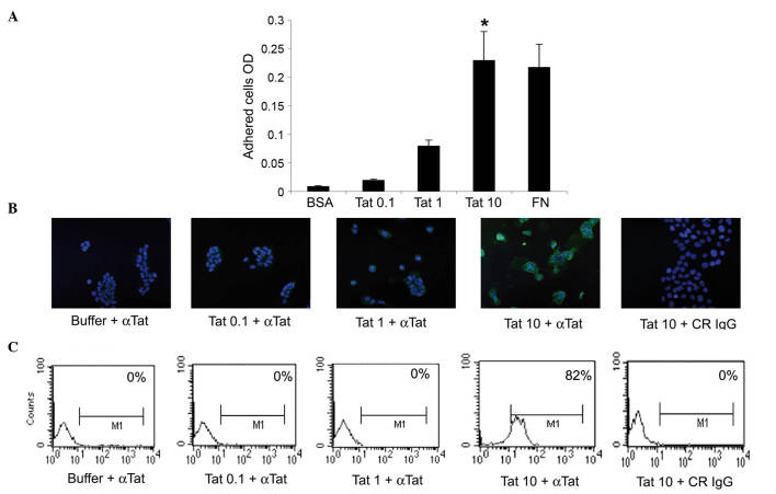
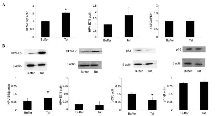
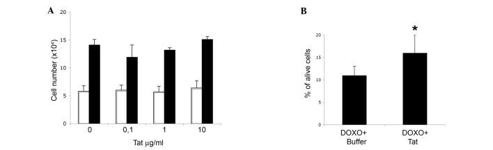
The infection of uterine cervical epithelial cells by oncogenic, high-risk human papilloma viruses (HR-HPVs) may lead to the development of cervical carcinoma. Of note, the incidence of this tumor is significantly increased in women infected by both HR-HPV and human immunodeficiency virus (HIV)-1. In this regard, previous studies have linked the HIV-1 Tat protein, a trans-activator of viral gene expression, to the pathogenesis of HIV-associated malignancies. In particular, it has been shown that upon its release by acutely infected cells, Tat protein can enter human cells, thus modifying their phenotype. Based on these findings, the present study evaluated whether extracellular Tat protein could be taken up by human uterine cervical carcinoma cells, and whether this could affect the expression of HPV (E6 or E7) or cellular (p16 or p53) molecules, which are key to cervical carcinoma development or progression. The results indicated that extracellular, biologically active HIV-1 Tat protein is taken up by human uterine cervical carcinoma cells, and that this is followed by an increase in the expression of the E6 protein of HPV, and by a reduction in the protein levels of the cellular oncosuppressor p53. Since p53 loss is associated with cell dedifferentiation and immortalization, these findings suggest a possible link between extracellular Tat protein and the high incidence and clinical aggressiveness of uterine cervical carcinoma observed in HIV/HPV doubly infected women.

Keywords: E6; HIV-1 Tat protein; human papilloma virus; p16; p53; uterine cervical carcinoma.


Figures

Figure 1.
Figure 1.
SiHa cells take up micromolar amounts of HIV-1 Tat protein. (A) SiHa cells were seeded on plates coated with 0.1, 1 or 10 µg/ml Tat. BSA or FN concentrations equimolar to 10 µg/ml Tat were employed as the negative or positive control, respectively. Adherent cells were quantified as described in the Materials and methods section. Results are expressed as mean optical density values from three experiments (*P<0.05). (B and C) SiHa cells were incubated for 30 min in medium containing 0.1, 1 or 10 µg/ml biologically active Tat or its suspension buffer (phosphate-buffered saline containing 0.1% BSA, which served as the negative control), and stained with anti-Tat or control antibodies. (B) Tat intracellular localization was visualized by fluorescence microscopy and photographed. Blue color corresponds to SiHa cell nuclei stained with 4′,6-diamidino-2-phenylindole, while green color indicated intracellular Tat protein, which was revealed as described in the Materials and methods section. Magnification, ×20. (C) The intracellular Tat content was evaluated by intracellular staining and flow cytometry. The percentage of positive cells (compared with isotype-stained samples) is reported in the boxes. The data in panels B and C correspond to a representative experiment. Repeated experiments produced similar results. OD, optical density; BSA, bovine serum albumin; FN, fibronectin; αTat, anti-Tat antibody; CR, control; IgG, immunoglublin G; M1, marker 1.
Figure 2.
Figure 2.
Uptake of extracellular Tat by SiHa cells is followed by a variation on HPV-E6 or p53 levels. SiHa cells were exposed for 48 h to 10 µg/ml Tat or its buffer. (A) Reverse transcription-quantitative polymerase chain reaction analysis of HPV-E6 (left panel), HPV-E7 (central panel) or p53 (right panel) RNA levels in SiHa cells cultured in the absence or presence of Tat. The results refer to the relative HPV-E6, HPV-E7 or p53 expression, normalized to the levels of β-actin or glyceraldehyde 3-phosphate dehydrogenase, which served as housekeeping genes. Bars represent the mean ± SD from four experiments (*P<0.05). (B) Cells were lysed, and equal amounts of total proteins were electrophoresed and analyzed by western blotting using a monoclonal antibody directed against the E6 or E7 protein of HPV16, human p53 or human p16ink4a. Blots were re-probed with anti-β-actin monoclonal antibody to verify equal loading of protein in each lane. The upper panels are representative western blots of HPV-E6, HPV-E7, p53, p16ink4a or β-actin. The lower panels represent the quantitative (densitometric) analysis of HPV-E6, HPV-E7, p53 or p16ink4a protein levels (normalized to those of β-actin) in control or Tat-treated SiHa cells. Bars represent the mean ± SD from 3–4 experiments (*P<0.05). SD, standard deviation; HPV, human papilloma virus; GAPDH, glyceraldehyde 3-phosphate dehydrogenase; INK4, inhibitor of cyclin-dependent kinase 4.
Figure 3.
Figure 3.
Entry of human immunodeficiency virus-1 Tat protein into SiHa cells partially rescues DOXO cytotoxic effect, with no impact on cell growth. SiHa cells were seeded at a density of 1.5×10⁴ cells/well in a 12-well cell culture plate. (A) Number of cells collected after 48 (white bars) or 96 h (black bars) of culture in medium containing 0.1, 1 or 10 µg/ml Tat, or its buffer (indicated as 0 µg/ml Tat). Data are the mean from two experiments, each performed in duplicate wells. (B) Cells were treated for 48 h with 10 µg/ml Tat or its buffer, and then grown for additional 48 h in the absence or presence of 2 µM DOXO. The results are expressed as the percentage of alive cells over the total cell number. Data represent the mean± standard deviation from three experiments (*P<0.05). DOXO, doxorubicin.

References

    1. Kang M, Cu-Uvin S. Association of HIV viral load and CD4 cell count with human papillomavirus detection and clearance in HIV-infected women initiating highly active antiretroviral therapy. HIV Med. 2012;13:372–378. doi: 10.1111/j.1468-1293.2011.00979.x. - DOI - PMC - PubMed
    1. Kim RH, Yochim JM, Kang MK, Shin KH, Christensen R, Park NH. HIV-1 Tat enhances replicative potential of human oral keratinocytes harboring HPV-16 genome. Int J Oncol. 2008;33:777–782. - PubMed
    1. Pol SB Vande, Klingelhutz AJ. Papillomavirus E6 oncoproteins. Virology. 2013;445:115–137. doi: 10.1016/j.virol.2013.04.026. - DOI - PMC - PubMed
    1. Shen R, Richter HE, Smith PD. Early HIV-1 target cells in human vaginal and ectocervical mucosa. Am J Reprod Immunol. 2011;65:261–267. doi: 10.1111/j.1600-0897.2010.00939.x. - DOI - PMC - PubMed
    1. Barillari G, Ensoli B. Angiogenic effects of extracellular HIV-1 Tat protein and its role in the pathogenesis of AIDS-associated Kaposi's sarcoma. Clin Microbiol Rev. 2002;15:310–326. doi: 10.1128/CMR.15.2.310-326.2002. - DOI - PMC - PubMed

LinkOut - more resources