

Ultrasonic features of papillary thyroid microcarcinoma coexisting with a thyroid abnormality
- PMID: 27698812
- PMCID: PMC5038495
- DOI: 10.3892/ol.2016.4999
Ultrasonic features of papillary thyroid microcarcinoma coexisting with a thyroid abnormality
Abstract
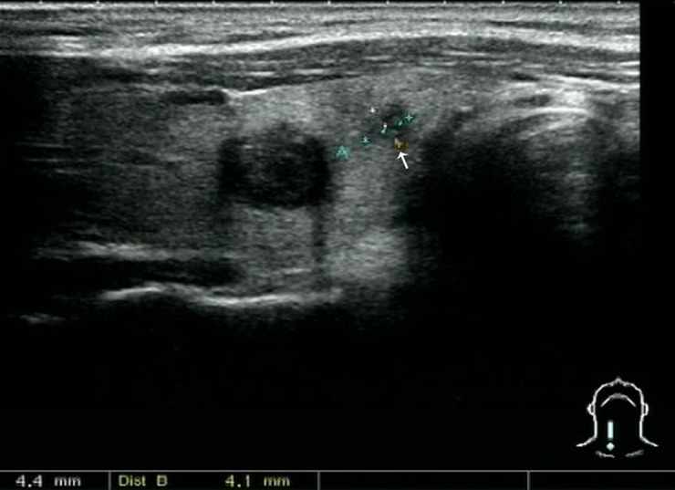
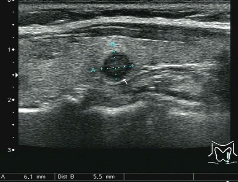
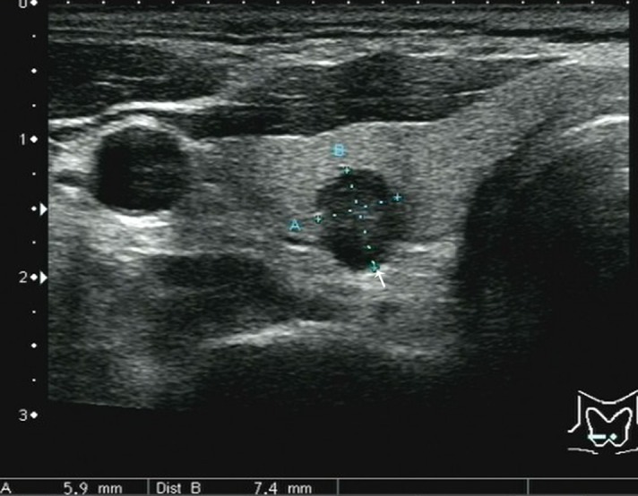
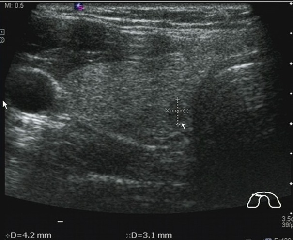
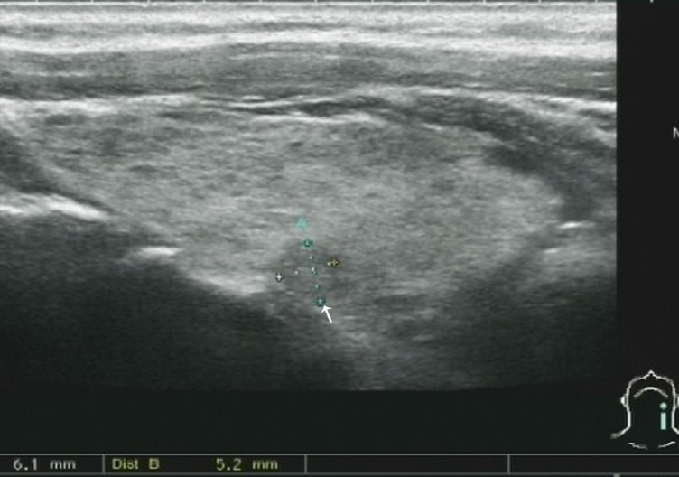
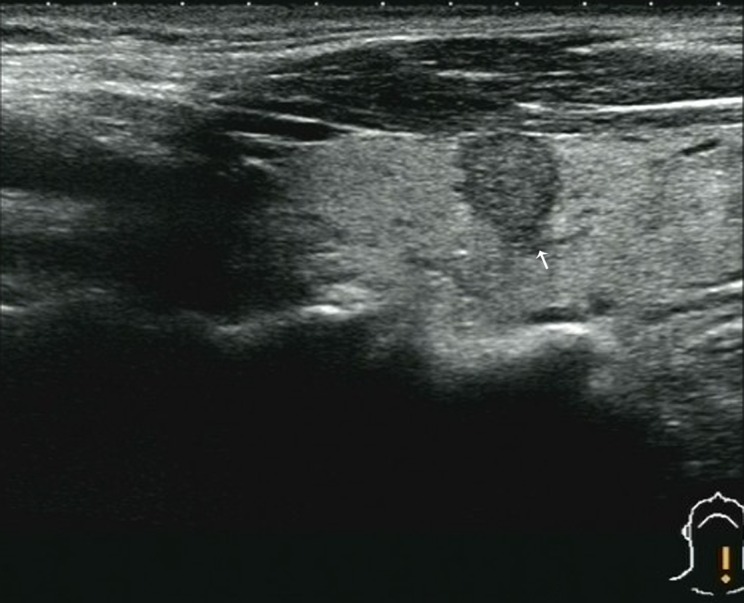
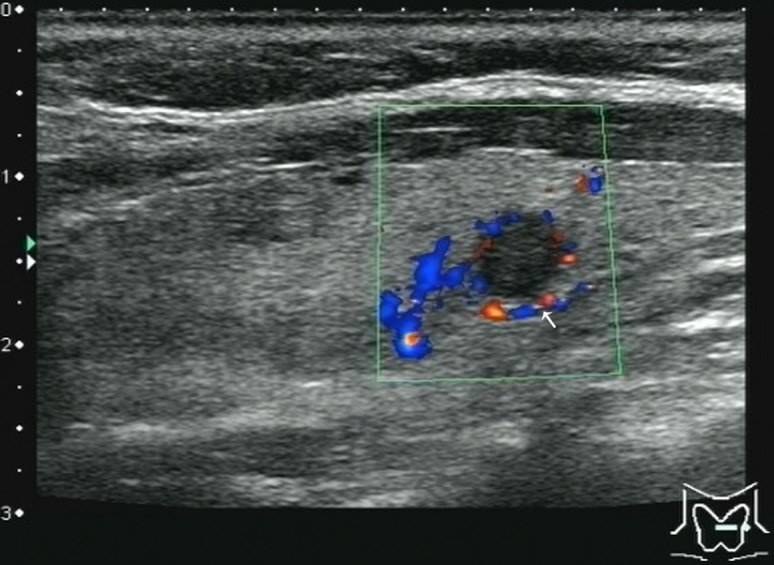
The present study aimed to investigate the value of ultrasonography in the diagnosis of papillary thyroid microcarcinoma (PTMC) coexisting with a thyroid abnormality, and to improve the accuracy of PTMC diagnosis. The ultrasonic features of 38 PTMC nodules coexisting with a thyroid abnormality and 56 thyroid benign nodules, obtained by surgical resection and confirmed by pathological analysis, were retrospectively analyzed. All masses were ≤ 1.0 cm in diameter. Ultrasonic features that were analyzed included the shape, aspect ratio, boundary, margin, echo, uniformity, presence or absence of microcalcification and enlargement of the lymph nodes, as well as the blood flow of the nodules. Furthermore, the sensitivity, specificity and accuracy of ultrasonography for the diagnosis of PTMC were obtained. The following ultrasonic features of thyroid nodules were significantly (P<0.05) associated with PTMC coexisting with a thyroid abnormality: An irregular shape; an aspect ratio of ≥ 1; an unclear boundary; blurred margins; internal heterogeneous hypoechogenicity; and microcalcification. Therefore, thyroid nodules with these ultrasonic characteristics coexisting with a thyroid abnormality may be suspected as malignant PTMC. The present study demonstrated that ultrasound-guided biopsies are necessary to prevent misdiagnosis of PTMC. The sensitivities of enlarged neck lymph nodes and abundant blood flow are so low that they may be considered as references for the differentiation of PTMC from benign nodules.
Keywords: papillary thyroid microcarcinoma; thyroid abnormality; ultrasonic diagnosis.
Figures
References
-
- Sugitani I, Kasai N, Fujimoto Y, Yanagisawa A. A novel classification system for patients with PTC: Addition of the new variables of large (3 cm or greater) nodal metastases and reclassification during the follow-up period. Surgery. 2004;135:139–148. doi: 10.1016/S0039-6060(03)00384-2. - DOI - PubMed
LinkOut - more resources
Full Text Sources
Other Literature Sources