

Interleukin-6 from Ovarian Mesenchymal Stem Cells Promotes Proliferation, Sphere and Colony Formation and Tumorigenesis of an Ovarian Cancer Cell Line SKOV3
- PMID: 27698921
- PMCID: PMC5039365
- DOI: 10.7150/jca.16116
Interleukin-6 from Ovarian Mesenchymal Stem Cells Promotes Proliferation, Sphere and Colony Formation and Tumorigenesis of an Ovarian Cancer Cell Line SKOV3
Abstract
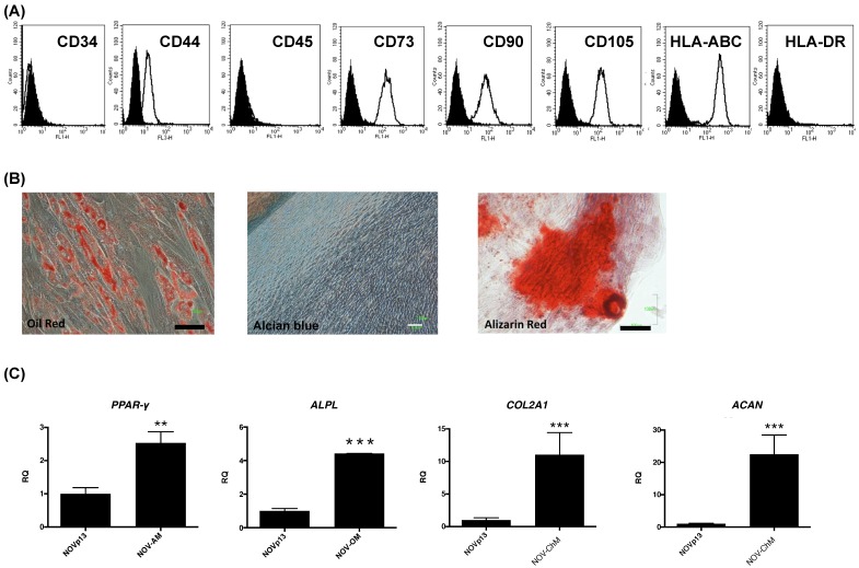
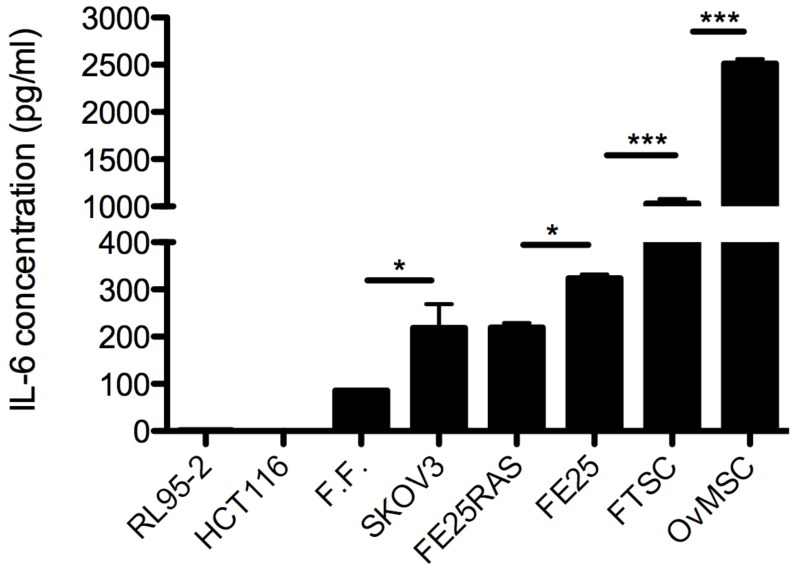
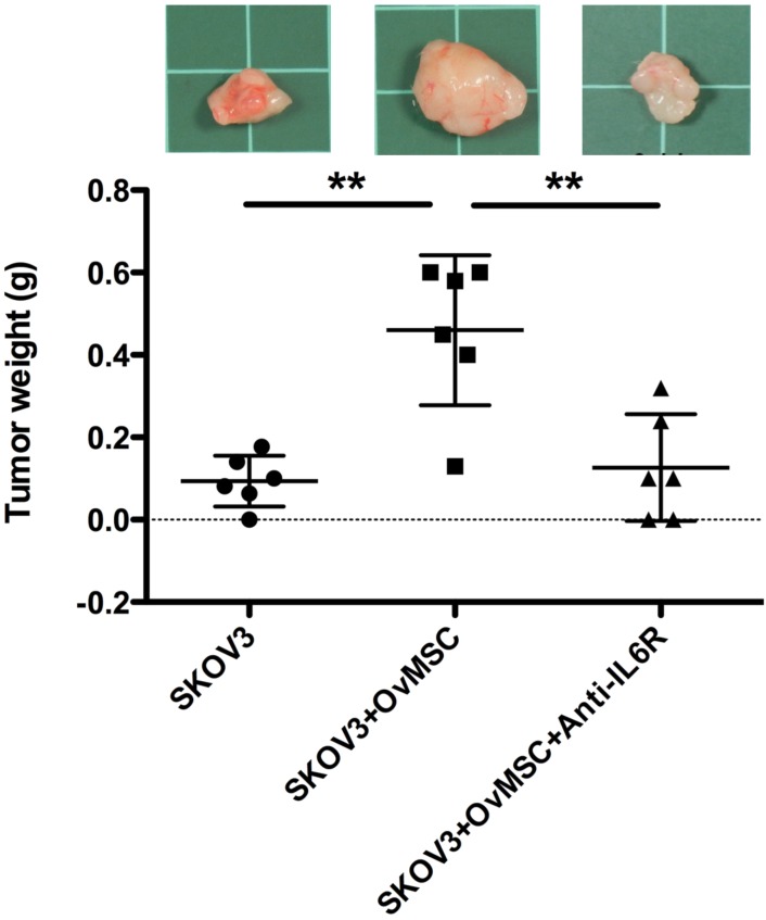
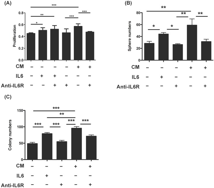
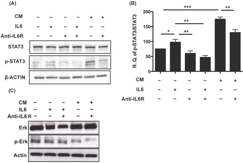
The origin of the majority of epithelial ovarian cancers (EOC) is regarded as extraovarian, with the ovary being the secondary site. The aim of this study was to explore the possible role of ovarian mesenchymal stem cells (OvMSCs) and secreted IL-6 in the development of EOC. OvMSCs were derived from normal ovarian stroma. Cell surface markers and differentiation capability were determined. The effects of IL-6 and conditioned medium of OvMSCs on the malignant phenotype of SKOV3 ovarian cancer cells were tested, and the status of STAT3 and ERK phosphorylation was investigated. OvMSCs had similar surface marker profiles as bone marrow mesenchymal stem cells, i.e., CD44 (+), CD90 (+) and CD45 (-), and was readily inducible to osteogenic, adipogenic and chondrogenic differentiation. OvMSCs secreted an extremely high level (>2500 pg/ml) of IL-6. Treatment of SKOV3 cells with conditioned media from OvMSCs increased cell proliferation, tumor sphere formation and anchorage independent growth, and resulted in activation of STAT3 but not ERK. Coinjection of OvMSCs with SKOV3 cell enhanced tumorigenesis in NOD-SCID mice. All of these behaviors were blocked by IL-6 receptor blocking antibody administered in vitro or in vivo. The OvMSCs alone injected into mice had no tumor growth after 3 months. By secreting high levels of IL-6, OvMSCs enhance the proliferation, sphere and colony formation and tumorigenesis of SKOV3 cells.
Keywords: IL-6, proliferation; SKOV3.; STAT3; tumorigenesis.
Conflict of interest statement
The authors have declared that no competing interest exists.
Figures
Similar articles
-
Co-culture of ovarian cancer stem-like cells with macrophages induced SKOV3 cells stemness via IL-8/STAT3 signaling.Biomed Pharmacother. 2018 Jul;103:262-271. doi: 10.1016/j.biopha.2018.04.022. Epub 2018 Apr 24. Biomed Pharmacother. 2018. PMID: 29656182
-
Bone Marrow Mesenchymal Stem Cells Promote Ovarian Cancer Cell Proliferation via Cytokine Interactions.Int J Mol Sci. 2024 Jun 19;25(12):6746. doi: 10.3390/ijms25126746. Int J Mol Sci. 2024. PMID: 38928452 Free PMC article.
-
[Effects of chrysin on sphere formation and CK2α expression of ovarian cancer stem-like cells derived from SKOV3 cell line].Zhonghua Yi Xue Za Zhi. 2016 Jul 5;96(25):2013-6. doi: 10.3760/cma.j.issn.0376-2491.2016.25.012. Zhonghua Yi Xue Za Zhi. 2016. PMID: 27470961 Chinese.
-
Inhibitory effects of metformin at low concentration on epithelial-mesenchymal transition of CD44(+)CD117(+) ovarian cancer stem cells.Stem Cell Res Ther. 2015 Dec 30;6:262. doi: 10.1186/s13287-015-0249-0. Stem Cell Res Ther. 2015. PMID: 26718286 Free PMC article.
-
Crosstalk between bone marrow-derived myofibroblasts and gastric cancer cells regulates cancer stemness and promotes tumorigenesis.Oncogene. 2016 Oct 13;35(41):5388-5399. doi: 10.1038/onc.2016.76. Epub 2016 Apr 25. Oncogene. 2016. PMID: 27109105 Free PMC article.
Cited by
-
Spontaneous Transformation of a p53 and Rb-Defective Human Fallopian Tube Epithelial Cell Line after Long Passage with Features of High-Grade Serous Carcinoma.Int J Mol Sci. 2022 Nov 10;23(22):13843. doi: 10.3390/ijms232213843. Int J Mol Sci. 2022. PMID: 36430324 Free PMC article.
-
Carcinoma-Associated Mesenchymal Stem Cells Promote Chemoresistance in Ovarian Cancer Stem Cells via PDGF Signaling.Cancers (Basel). 2020 Jul 27;12(8):2063. doi: 10.3390/cancers12082063. Cancers (Basel). 2020. PMID: 32726910 Free PMC article.
-
Leukemia inhibitory factor functions in parallel with interleukin-6 to promote ovarian cancer growth.Oncogene. 2019 Feb;38(9):1576-1584. doi: 10.1038/s41388-018-0523-6. Epub 2018 Oct 10. Oncogene. 2019. PMID: 30305729 Free PMC article.
-
Crosstalk Between Mesenchymal Stromal Cells and Tumor-Associated Macrophages in Gastric Cancer.Front Oncol. 2020 Oct 9;10:571516. doi: 10.3389/fonc.2020.571516. eCollection 2020. Front Oncol. 2020. PMID: 33163402 Free PMC article. Review.
-
Emerging role of mesenchymal stromal cells in gynecologic cancer therapy.Stem Cell Res Ther. 2023 Dec 5;14(1):347. doi: 10.1186/s13287-023-03585-0. Stem Cell Res Ther. 2023. PMID: 38049868 Free PMC article. Review.
References
-
- Siegel R, Ward E, Brawley O. et al. Cancer statistics, 2011: the impact of eliminating socioeconomic and racial disparities on premature cancer deaths. CA Cancer J Clin. 2011;61(4):212–236. - PubMed
-
- Lim D, Oliva E. Precursors and pathogenesis of ovarian carcinoma. Pathology. 2013;45(3):229–242. - PubMed
-
- Huang Y, Lin Z, Zhuang J. et al. Prognostic significance of alpha V integrin and VEGF in osteosarcoma after chemotherapy. Onkologie. 2008;31(10):535–540. - PubMed
LinkOut - more resources
Full Text Sources
Other Literature Sources
Research Materials
Miscellaneous
