

Senescence Process in Primary Wilms' Tumor Cell Culture Induced by p53 Independent p21 Expression
- PMID: 27698927
- PMCID: PMC5039371
- DOI: 10.7150/jca.16316
Senescence Process in Primary Wilms' Tumor Cell Culture Induced by p53 Independent p21 Expression
Abstract
Wilms tumor (WT) is an embryonal tumor occurring in developing kidney tissue. WT cells showing invasive cancer characteristics, also retain renal stem cell behaviours. In-vitro culture of WT is hampered by limited replicative potential. This study aimed to establish a longterm culture of WT cells to enable the study of molecular events to attempt to explain its cellular senescence.
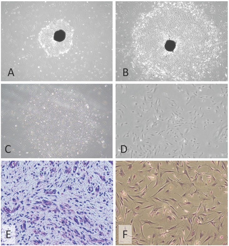
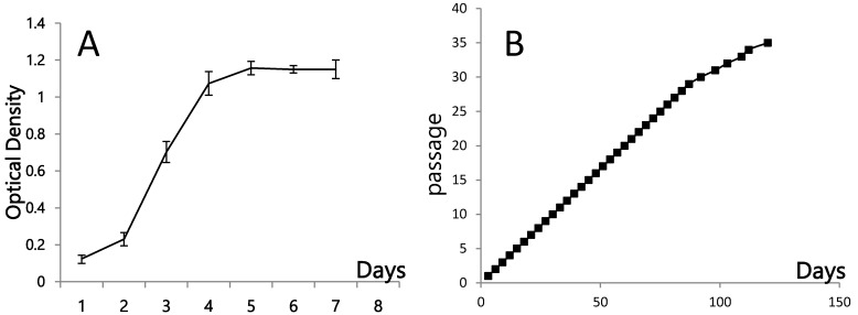
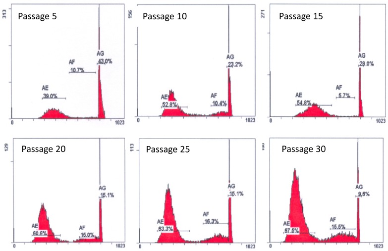
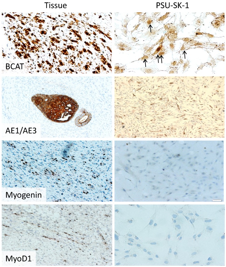
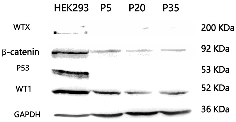
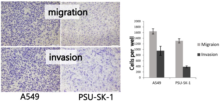
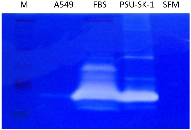
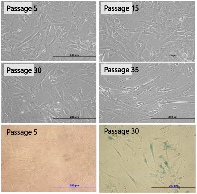
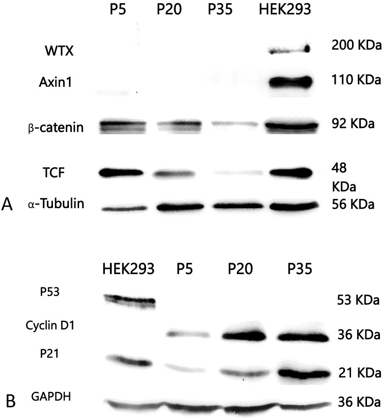
Methods: Primary cell cultures from fresh WT tumor specimen were established. Of 5 cultures tried, only 1 could be propagated for more than 7 passages. One culture, identified as PSU-SK-1, could be maintained > 35 passages and was then subjected to molecular characterization and evaluation for cancer characteristics. The cells consistently harbored concomitant mutations of CTNNB1 (Ser45Pro) and WT1 (Arg413Stop) thorough the cultivation. On Transwell invasion assays, the cells exhibited migration and invasion at 55% and 27% capability of the lung cancer cells, A549. On gelatin zymography, PSU-SK-1 showed high expression of the matrix metaloproteinase. The cells exhibited continuous proliferation with 24-hour doubling time until passages 28-30 when the growth slowed, showing increased cell size, retention of cells in G1/S proportion and positive β-galactosidase staining. As with those evidence of senescence in advanced cell passages, expression of p21 and cyclin D1 increased when the expression of β-catenin and its downstream protein, TCF, declined. There was also loss-of-expression of p53 in this cell line. In conclusion, cellular senescence was responsible for limited proliferation in the primary culture of WT, which was also associated with increased expression of p21 and was independent of p53 expression. Decreased activation of the Wnt signalling might explain the induction of p21 expression.
Keywords: Primary cell culture; Senescence.; Wilms tumor.
Conflict of interest statement
The authors have declared that no competing interest exists.
Figures
Similar articles
-
Wilms tumor cells with WT1 mutations have characteristic features of mesenchymal stem cells and express molecular markers of paraxial mesoderm.Hum Mol Genet. 2010 May 1;19(9):1651-68. doi: 10.1093/hmg/ddq042. Epub 2010 Jan 27. Hum Mol Genet. 2010. PMID: 20106868
-
CTNNB1 mutations and overexpression of Wnt/beta-catenin target genes in WT1-mutant Wilms' tumors.Am J Pathol. 2004 Dec;165(6):1943-53. doi: 10.1016/s0002-9440(10)63246-4. Am J Pathol. 2004. PMID: 15579438 Free PMC article.
-
Single-cell analysis of p16(INK4a) and p21(WAF1) expression suggests distinct mechanisms of senescence in normal human and Li-Fraumeni Syndrome fibroblasts.J Cell Physiol. 2010 Apr;223(1):57-67. doi: 10.1002/jcp.22002. J Cell Physiol. 2010. PMID: 20039273
-
In vitro lifespan and senescence mechanisms of human nucleus pulposus chondrocytes.Spine J. 2014 Mar 1;14(3):499-504. doi: 10.1016/j.spinee.2013.06.099. Epub 2013 Dec 15. Spine J. 2014. PMID: 24345469
-
Nephroblastoma (Wilms' tumor): a model system of aberrant renal development.Semin Diagn Pathol. 1994 May;11(2):126-35. Semin Diagn Pathol. 1994. PMID: 7809506 Review.
Cited by
-
Competitive endogenous RNA (ceRNA) regulation network of lncRNAs, miRNAs, and mRNAs in Wilms tumour.BMC Med Genomics. 2019 Dec 16;12(1):194. doi: 10.1186/s12920-019-0644-y. BMC Med Genomics. 2019. PMID: 31842887 Free PMC article.
-
Comprehensive Biology and Genetics Compendium of Wilms Tumor Cell Lines with Different WT1 Mutations.Cancers (Basel). 2020 Dec 28;13(1):60. doi: 10.3390/cancers13010060. Cancers (Basel). 2020. PMID: 33379206 Free PMC article.
-
Overexpression of CCDC69 activates p14ARF/MDM2/p53 pathway and confers cisplatin sensitivity.J Ovarian Res. 2019 Jan 16;12(1):4. doi: 10.1186/s13048-019-0479-3. J Ovarian Res. 2019. PMID: 30651135 Free PMC article.
-
Management and potentialities of primary cancer cultures in preclinical and translational studies.J Transl Med. 2017 Nov 7;15(1):229. doi: 10.1186/s12967-017-1328-z. J Transl Med. 2017. PMID: 29116016 Free PMC article. Review.
-
Wilms' Tumor Primary Cells Display Potent Immunoregulatory Properties on NK Cells and Macrophages.Cancers (Basel). 2021 Jan 9;13(2):224. doi: 10.3390/cancers13020224. Cancers (Basel). 2021. PMID: 33435455 Free PMC article.
References
LinkOut - more resources
Full Text Sources
Other Literature Sources
Research Materials
Miscellaneous
