Brain Formaldehyde is Related to Water Intake behavior
- PMID: 27699080
- PMCID: PMC5036952
- DOI: 10.14336/AD.2016.0323
Brain Formaldehyde is Related to Water Intake behavior
Abstract
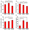
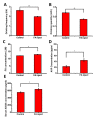
A promising strategy for the prevention of Alzheimer's disease (AD) is the identification of age-related changes that place the brain at risk for the disease. Additionally, AD is associated with chronic dehydration, and one of the significant changes that are known to result in metabolic dysfunction is an increase in the endogenous formaldehyde (FA) level. Here, we demonstrate that the levels of uric formaldehyde in AD patients were markedly increased compared with normal controls. The brain formaldehyde levels of wild-type C57 BL/6 mice increased with age, and these increases were followed by decreases in their drinking frequency and water intake. The serum arginine vasopressin (AVP) concentrations were also maintained at a high level in the 10-month-old mice. An intravenous injection of AVP into the tail induced decreases in the drinking frequency and water intake in the mice, and these decreases were associated with increases in brain formaldehyde levels. An ELISA assay revealed that the AVP injection increased both the protein level and the enzymatic activity of semicarbazide-sensitive amine oxidase (SSAO), which is an enzyme that produces formaldehyde. In contrast, the intraperitoneal injection of formaldehyde increased the serum AVP level by increasing the angiotensin II (ANG II) level, and this change was associated with a marked decrease in water intake behavior. These data suggest that the interaction between formaldehyde and AVP affects the water intake behaviors of mice. Furthermore, the highest concentration of formaldehyde in vivo was observed in the morning. Regular water intake is conducive to eliminating endogenous formaldehyde from the human body, particularly when water is consumed in the morning. Establishing good water intake habits not only effectively eliminates excess formaldehyde and other metabolic products but is also expected to yield valuable approaches to reducing the risk of AD prior to the onset of the disease.
Keywords: arginine vasopressin; behavior; formaldehyde; semicarbazide-sensitive amine oxidase; water intake.
Figures


Comment in
-
Depression, Diabetes and Dementia: Formaldehyde May Be a Common Causal Agent; Could Carnosine, a Pluripotent Peptide, Be Protective?Aging Dis. 2017 Apr 1;8(2):128-130. doi: 10.14336/AD.2017.0120. eCollection 2017 Apr. Aging Dis. 2017. PMID: 28400979 Free PMC article. No abstract available.
References
-
- Luckey AE, Parsa CJ (2003). Fluid and electrolytes in the aged. Archives of Surgery, 138(10): 1055-1060 - PubMed
-
- Stout NR, Kenny RA, Baylis PH (1999). A review of water balance in ageing in health and disease Gerontology, 45(2): 61-66 - PubMed
-
- Purdy M (2006). Accelerating weight loss may signal development of Alzheimer’s disease. Bio-Medicine, http://news.bio-medicine.org/medicine-news-3/Accelerating-weight-loss-ma...
-
- Gharibzadeh Hoseini (2007).Chronic dehydration may be a preventable risk factor for Alzheimer’s disease. Med Hypoth, 68(3): 718 - PubMed
-
- Buffa R, Mereu RM, Putzu PF, Floris G, Marini E (2010). Bioelectrical impedance vector analysis detects low body cell mass and dehydration in the patients with Alzheimer’s disease. JNHA, 14(10): 823-874 - PubMed
LinkOut - more resources
Full Text Sources
Other Literature Sources
Miscellaneous
