

Immunologic factors may play a role in herpes simplex virus 1 reactivation in the brain and retina after influenza vaccination
- PMID: 27699152
- PMCID: PMC5045948
- DOI: 10.1016/j.idcr.2016.09.012
Immunologic factors may play a role in herpes simplex virus 1 reactivation in the brain and retina after influenza vaccination
Abstract
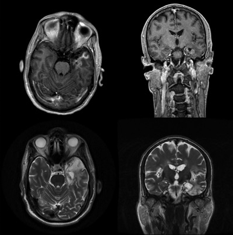
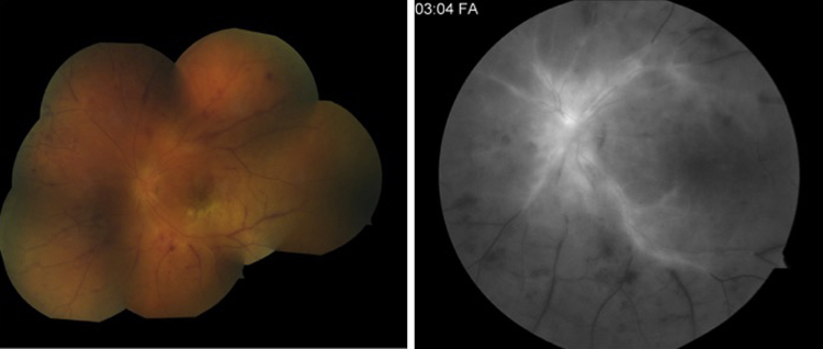
Herpes simplex virus 1 (HSV-1) is a nearly ubiquitous human pathogen, remaining dormant in its human host the majority of the time. The interaction between HSV-1 and the immune system represents a complicated balance of power that allows the virus to persist in the host for a lifetime. However, disruptions in the immune system can activate the virus with the potential to cause devastating infections in the central nervous system (CNS). We present a patient who suffered three consecutive yearly HSV-1 CNS episodes (encephalitis, seizure, and retinitis), each within days of his influenza vaccination. We highlight subtle immunologic defects in this patient that may have allowed unchecked viral replication and resultant disease manifestations, as well as the potential role of influenza vaccine in tipping this balance in favor of HSV-1.
Keywords: Acute retinal necrosis; Encephalitis; Herpes simplex; Influenza; Vaccination.
Figures
References
-
- Motani H., Sakurada K., Ikegaya H., Akutsu T., Hayakawa M., Sato Y. Detection of herpes simplex virus type 1 DNA in bilateral human trigeminal ganglia and optic nerves by polymerase chain reaction. J Med Virol. 2006;78(12):1584–1587. - PubMed
-
- Steiner I., Mador N., Reibstein I., Spivack J.G., Fraser N.W. Herpes simplex virus type 1 gene expression and reactivation of latent infection in the central nervous system. Neuropathol Appl Neurobiol. 1994;20(3):253–260. - PubMed
Publication types
LinkOut - more resources
Full Text Sources
Other Literature Sources
