. 2016 Aug 18;1(13):e84671.
doi: 10.1172/jci.insight.84671.

Quantum coherence spectroscopy to measure dietary fat retention in the liver

Affiliations

Quantum coherence spectroscopy to measure dietary fat retention in the liver

Lucas Lindeboom et al. JCI Insight. .

Abstract

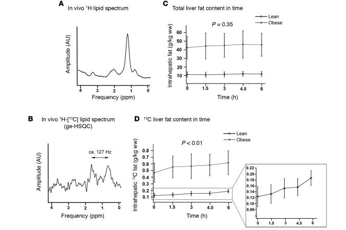
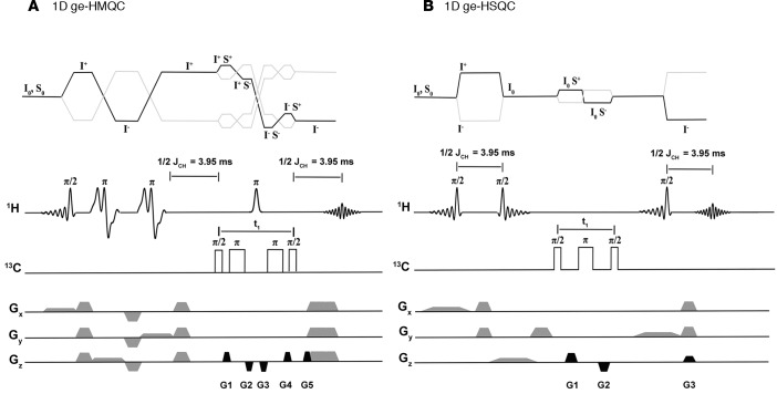
The prevalence of fatty liver reaches alarming proportions. Fatty liver increases the risk for insulin resistance, cardiovascular disease, and nonalcoholic steatohepatitis (NASH). Although extensively studied in a preclinical setting, the lack of noninvasive methodologies hampers our understanding of which pathways promote hepatic fat accumulation in humans. Dietary fat retention is one of the pathways that may lead to fatty liver. The low (1.1%) natural abundance (NA) of carbon-13 (13C) allows use of 13C-enriched lipids for in vivo MR studies. Successful implementation of such methodology, however, is challenging due to low sensitivity of 13C-magnetic resonance spectroscopy (13C-MRS). Here, we investigated the use of 1-dimensional gradient enhanced heteronuclear single quantum coherence (ge-HSQC) spectroscopy for the in vivo detection of hepatic 1H-[13C]-lipid signals after a single high-fat meal with 13C-labeled fatty acids in 5 lean and 6 obese subjects. Postprandial retention of orally administered 13C-labeled fatty acids was significant (P < 0.01). Approximately 1.5% of the tracer was retained in the liver after 6 hours, and retention was similar in both groups (P = 0.92). Thus, a substantial part of the liver fat can originate directly from storage of meal-derived fat. The ge-HSQC can be used to noninvasively reveal the contribution of dietary fat to the development of hepatic steatosis over time.


Figures

Figure 1
Figure 1. Spectra acquired from the phantom containing naturally abundant 13C Intralipid.
(A and B) The result from the conventional quantum coherence sequences. In both cases, the 13C chemical shift evolution during the t1 period resulted in phase distortions in the spectrum. Addition of the 13C inversion pulse led to nonphase distorted lipid spectra, in which both CH2 and CH3 signals were observable. (C and D) Both relative and absolute signals were higher in the 1D ge-HSQC (heteronuclear single quantum coherence) as compared with the 1D ge-HMQC (heteronuclear multiple quantum coherence).
Figure 2
Figure 2. Spectra acquired from the Intralipid phantom with increasing t1 in the 1D ge-HMQC (heteronuclear multiple quantum coherence) and the 1D ge-HSQC (heteronuclear single quantum coherence).
(A and B) In A, the results for the 1D ge-HMQC are shown, while in B, the outcome of the 1D ge-HSQC sequence is presented. The evolution time (t1) was increased with 1 ms every step and started at, respectively, 7.6 and 4.2 ms. It is apparent that the signal acquired with the ge-HMQC decays rapidly with an increasing t1.
Figure 3
Figure 3. Spectra acquired from the phantom with the uniformly labeled 13C tracer with increasing t1 in the 1D ge-HSQC (heteronuclear single quantum coherence).
The evolution time (t1) was increased with 1 ms every step and started at 4.2 ms. Due to the 13C-13C couplings in the tracer, the signal now decays rapidly with increasing t1.
Figure 4
Figure 4. Results from the in vivo experiments.
(A and B) Typical example of the total lipid spectrum, acquired with STEAM (stimulated echo acquistion mode) 1H-MRS, and the concomitant 13C-edited lipid spectrum, acquired with the ge-HSQC (heteronuclear single quantum coherence) sequence. As no decoupling was applied, two lipid CH2 peaks are visible, which are separated by approximately 127 Hz. (C) The total IHL (intrahepatic lipid) pool (g/kg ww) for both lean (n = 5) and obese (n = 6) subjects at 1,5, 3.0, 4.5, and 6.0 hours after a high-fat meal. Total IHL did not change after the meal (repeated-measures ANOVA, P = 0.35). (D) The absolute concentration of 13C lipids (g/kg ww) in the liver is plotted in time, showing retention of 13C-labeled fatty acids after the meal (repeated-measures ANOVA, P < 0.01).
Figure 5
Figure 5. Pulse sequences used in this study.
(A) The 1D ge-HMQC (heteronuclear multiple quantum coherence) sequence is shown. The HMQC block was placed after PRESS (point resolved spectroscopy) localization. Two additional 13C inversion pulses were used to refocus the 13C chemical shift evolution. Minimum evolution time (t1) was 7.6 ms. In contrast to the conventional ge-HMQC sequence, 5 gradients were used for coherence selection. Gradients were applied in a ratio of 1:–1:–1:1:1. (B) The 1D ge-HSQC (heteronuclear single quantum coherence) sequence, based on STEAM (stimulated echo acquistion mode) localization. The inversion pulse during the t1, which was used on the 1H channel previously, was now applied on the 13C channel to again refocus 13C chemical shift evolution. Minimum t1 was 4.2 ms, and gradients were applied in a 2:–2:1 ratio. In both sequences, 1/2JCH was chosen as 3.95 (J = 127 Hz for CH2 lipids). No decoupling was applied.

References

    1. Szczepaniak LS, et al. Measurement of intracellular triglyceride stores by H spectroscopy: validation in vivo. Am J Physiol. 1999;276(5 Pt 1):E977–E989. - PubMed
    1. Boesch C, Slotboom J, Hoppeler H, Kreis R. In vivo determination of intra-myocellular lipids in human muscle by means of localized 1H-MR-spectroscopy. Magn Reson Med. 1997;37(4):484–493. doi: 10.1002/mrm.1910370403. - DOI - PubMed
    1. Hwang JH, et al. Increased intrahepatic triglyceride is associated with peripheral insulin resistance: in vivo MR imaging and spectroscopy studies. Am J Physiol Endocrinol Metab. 2007;293(6):E1663–E1669. doi: 10.1152/ajpendo.00590.2006. - DOI - PubMed
    1. Korenblat KM, Fabbrini E, Mohammed BS, Klein S. Liver, muscle, and adipose tissue insulin action is directly related to intrahepatic triglyceride content in obese subjects. Gastroenterology. 2008;134(5):1369–1375. doi: 10.1053/j.gastro.2008.01.075. - DOI - PMC - PubMed
    1. Seppälä-Lindroos A, et al. Fat accumulation in the liver is associated with defects in insulin suppression of glucose production and serum free fatty acids independent of obesity in normal men. J Clin Endocrinol Metab. 2002;87(7):3023–3028. - PubMed

Publication types