. 2016 Sep 8;1(14):e86017.
doi: 10.1172/jci.insight.86017.

The antifibrotic drug pirfenidone promotes pulmonary cavitation and drug resistance in a mouse model of chronic tuberculosis

Affiliations

The antifibrotic drug pirfenidone promotes pulmonary cavitation and drug resistance in a mouse model of chronic tuberculosis

Bintou A Ahidjo et al. JCI Insight. .

Abstract

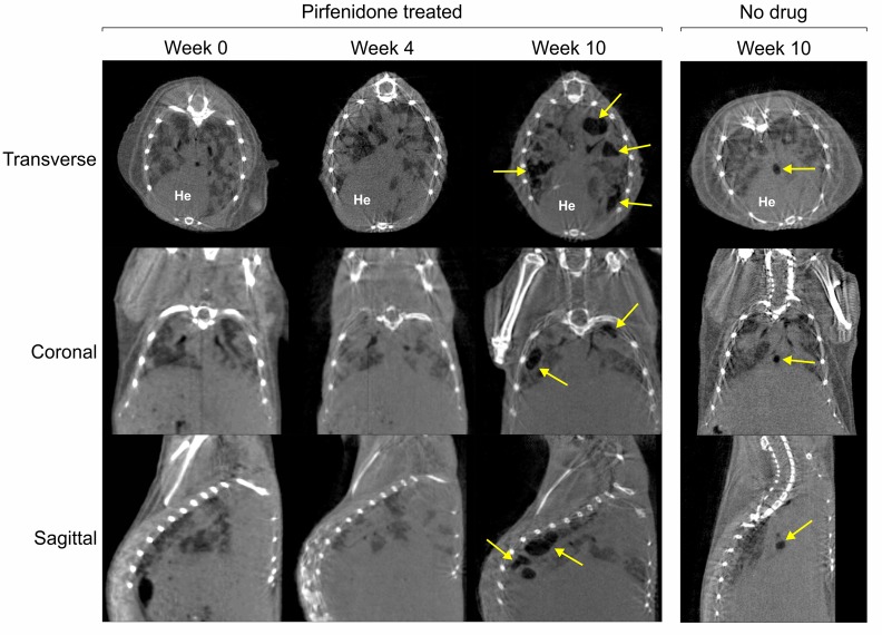
Pirfenidone is a recently approved antifibrotic drug for the treatment of idiopathic pulmonary fibrosis (IPF). Because tuberculosis (TB) is characterized by granulomatous inflammation in conjunction with parenchymal destruction and replacement fibrosis, we sought to determine whether the addition of pirfenidone as an adjunctive, host-directed therapy provides a beneficial effect during antimicrobial treatment of TB. We hypothesized that pirfenidone's antiinflammatory and antifibrotic properties would reduce inflammatory lung damage and increase antimicrobial drug penetration in granulomas to accelerate treatment response. The effectiveness of adjunctive pirfenidone during TB drug therapy was evaluated using a murine model of chronic TB. Mice treated with standard therapy 2HRZ/4HR (H, isoniazid; R, rifampin; and Z, pyrazinamide) were compared with 2 alternative regimens containing pirfenidone (Pf) (2HRZPf/4HRPf and 2HRZPf/4HR). Contrary to our hypothesis, adjunctive pirfenidone use leads to reduced bacterial clearance and increased relapse rates. This treatment failure is closely associated with the emergence of isoniazid monoresistant bacilli, increased cavitation, and significant lung pathology. While antifibrotic agents may eventually be used as part of adjunctive host-directed therapy of TB, this study clearly demonstrates that caution must be exercised. Moreover, as pirfenidone becomes more widely used in clinical practice, increased patient monitoring would be required in endemic TB settings.


Figures

Figure 1
Figure 1. Effect of pirfenidone on Mtb-infected C3HeB/FeJ mice as assessed by MMP levels, cytokine levels, and colony forming units.
(A) MMPSense 680, a fluorescent in vivo imaging agent activated by MMPs, was used to assess MMP activity in n ≥ 2 mice per group. As per the manufacturer’s experimental protocol, the higher the intensity the greater the MMP activity. (B) Lung cytokines assessed by ELISA represent mean (± SD) from n ≥ 3 mice per group. For comparison between treatment groups, cytokines were analyzed by 2-way ANOVA with post-hoc Tukey honest significant difference (HSD) tests. For comparison within treatment groups, cytokines were analyzed by 1-way ANOVA with post-hoc Sidak tests. All measures of variations are expressed as ± SD. (C) Lung colony forming units (CFU); data represent the mean ± SD from n ≥ 5 mice. Lung CFU counts (x) were log-transformed as log10 (x + 1) prior to analysis and 1-way ANOVA tests used to determine statistical significance between the groups. Pf, pirfenidone; ND, not determined. *P < 0.05.
Figure 2
Figure 2. Efficacy of pirfenidone as an adjunct for tuberculosis therapy in C3HeB/FeJ mice as assessed by CFU, relapse rates, and gross pathology.
(A) All lung CFU data represent the mean ± SD from n ≥ 3 mice. (BE) Each data point represents a single mouse. Red symbols indicate mice that harbored isoniazid monoresistant bacilli (CE). All lung CFU counts (x) were log-transformed as log10 (x + 1) prior to analysis and 1-way ANOVA tests used to determine statistical significance of differences between the groups. All measures of variations are expressed as ± SD. Relapse rates (F) of n ≥ 7 mice at each time point were compared using Fisher’s exact test. (G) Representative mouse lung out of n ≥ 20 mice receiving 2 months standard therapy. (H) Representative mouse lung out of n ≥ 20 mice receiving 2 months standard therapy with pirfenidone. UN, untreated; H, isoniazid; R, rifampin; Z, pyrazinamide; Pf, pirfenidone.
Figure 3
Figure 3. Experimental scheme 3.
Eleven control mice and 14 test mice were infected with Mycobacterium tuberculosis (Mtb). Weeks 0–4 indicate early treatment and Weeks 4–10 indicate late treatment times. H, isoniazid; R, rifampin; Pf, pirfenidone
Figure 4
Figure 4. Pathology of Mycobacterium tuberculosis (Mtb)–infected C3HeB/FeJ mice in the absence and presence of pirfenidone (Pf).
(A) Histopathology of lungs of Mtb-infected C3HeB/FeJ Pf-naive mice showing reduced MMPsense 680 fluorescence (left) and hence reduced MMP activation, areas of hypercellularity, and a single large necrogranuloma (G), composed of a central core of necrotic granular debris (N), lined by foamy macrophages (FM) and surrounded by a rim of fibrous connective tissue. Middle 4 images are of H&E-stained tissue. Scale bars: 200 μm. Images on right are of acid-fast–stained tissue. Scale bars: 50 μm. (B) Pf treatment resulted in increased MMPsense 680 fluorescence, which corresponds to an increase in MMP activity and an increased number of pulmonary cavities with air-filled centers (*), lined by foamy macrophages (FM) and surrounded by a thin rim of fibrous connective tissue (CT). Cavities are interspersed with adjacent normal alveoli (A). (C) Gross pathology of lobar pneumonia observed during treatment with pirfenidone (red arrows). These are all representative of n ≥ 3 characterized mouse lungs per group.
Figure 5
Figure 5. Imaging reveals increased cavitation during pirfenidone treatment.
Transverse, coronal, and sagittal sections of serial CT imaging performed on a Mycobacterium tuberculosis–infected mouse treated with pirfenidone (panels on the left) and untreated control (panel on the right). These are representative of 5 control mice receiving standard therapy and 4 mice receiving adjunctive pirfenidone treatment. Ten weeks after treatment, cavitary lesions of control mice progressively resolved, while in pirfenidone-treated mice multiple progressive cavitary lesions can be seen in the lung parenchyma (yellow arrows) despite standard treatment. Tuberculous lesions are visible as radiodense areas on the CT images. He, heart.
Figure 6
Figure 6. Disease severity of lung sections obtained from at least 3 mice before (early) and after (late) antimycobacterial treatment.
All graphs shown as median and interquartile range. All values are P > 0.05. Here, 11 control mice and 14 test mice were infected with Mycobacterium tuberculosis. Early = the first 4 weeks after at the start of treatment; Late = 4–10 weeks from start of treatment. During the early phase of treatment, 5 control mice and 3 pirfendone-treated (Pf-treated) mice died and so 5 control mice and 11 Pf-treated mice were analyzed during the late treatment phase.

References

    1. Schaefer CJ, Ruhrmund DW, Pan L, Seiwert SD, Kossen K. Antifibrotic activities of pirfenidone in animal models. Eur Respir Rev. 2011;20(120):85–97. doi: 10.1183/09059180.00001111. - DOI - PMC - PubMed
    1. Noble PW, et al. Pirfenidone in patients with idiopathic pulmonary fibrosis (CAPACITY): two randomised trials. Lancet. 2011;377(9779):1760–1769. doi: 10.1016/S0140-6736(11)60405-4. - DOI - PubMed
    1. Taniguchi H, et al. Pirfenidone in idiopathic pulmonary fibrosis. Eur Respir J. 2010;35(4):821–829. doi: 10.1183/09031936.00005209. - DOI - PubMed
    1. Macías-Barragán J, Sandoval-Rodríguez A, Navarro-Partida J, Armendáriz-Borunda J. The multifaceted role of pirfenidone and its novel targets. Fibrogenesis Tissue Repair. 2010;3:e86017. - PMC - PubMed
    1. Editorial Fibrosis in tuberculosis. Journal of the American Medical Association. 1927;89 doi: 10.1001/jama.1927.02690200046016.

Publication types