. 2016 Mar 17;1(3):e85461.
doi: 10.1172/jci.insight.85461.

Identifying candidate genes for 2p15p16.1 microdeletion syndrome using clinical, genomic, and functional analysis

Affiliations

Identifying candidate genes for 2p15p16.1 microdeletion syndrome using clinical, genomic, and functional analysis

Hani Bagheri et al. JCI Insight. .

Abstract

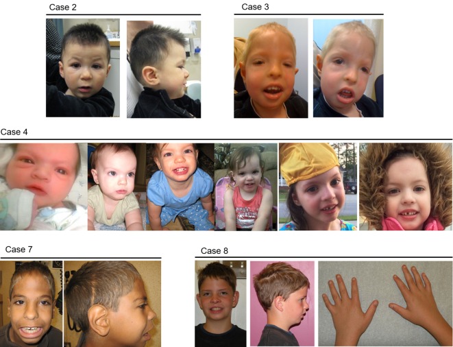
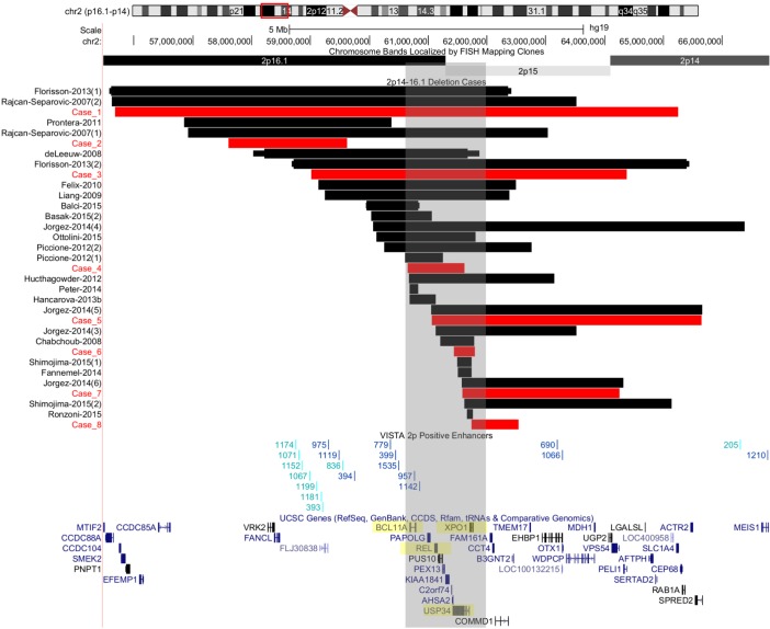
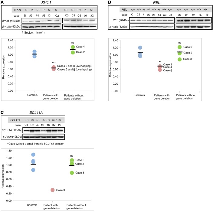
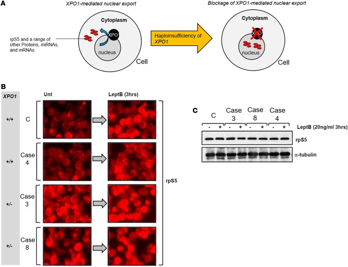
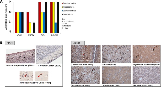
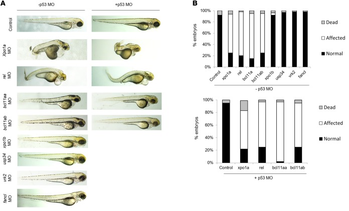
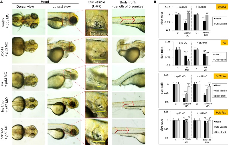
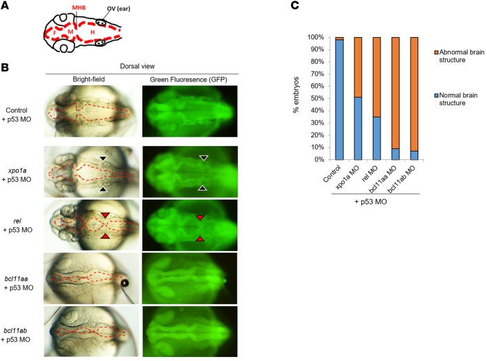
The 2p15p16.1 microdeletion syndrome has a core phenotype consisting of intellectual disability, microcephaly, hypotonia, delayed growth, common craniofacial features, and digital anomalies. So far, more than 20 cases of 2p15p16.1 microdeletion syndrome have been reported in the literature; however, the size of the deletions and their breakpoints vary, making it difficult to identify the candidate genes. Recent reports pointed to 4 genes (XPO1, USP34, BCL11A, and REL) that were included, alone or in combination, in the smallest deletions causing the syndrome. Here, we describe 8 new patients with the 2p15p16.1 deletion and review all published cases to date. We demonstrate functional deficits for the above 4 candidate genes using patients' lymphoblast cell lines (LCLs) and knockdown of their orthologs in zebrafish. All genes were dosage sensitive on the basis of reduced protein expression in LCLs. In addition, deletion of XPO1, a nuclear exporter, cosegregated with nuclear accumulation of one of its cargo molecules (rpS5) in patients' LCLs. Other pathways associated with these genes (e.g., NF-κB and Wnt signaling as well as the DNA damage response) were not impaired in patients' LCLs. Knockdown of xpo1a, rel, bcl11aa, and bcl11ab resulted in abnormal zebrafish embryonic development including microcephaly, dysmorphic body, hindered growth, and small fins as well as structural brain abnormalities. Our multifaceted analysis strongly implicates XPO1, REL, and BCL11A as candidate genes for 2p15p16.1 microdeletion syndrome.


Figures

Figure 1
Figure 1. Physical features of the 2p15p16.1 microdeletion carriers.
Images of the patients in cases 2, 3, 4, 7, and 8 illustrating the craniofacial and digital abnormalities seen in 2p15p16.1 microdeletion syndrome. All patients had microcephaly and/or head shape abnormalities (except case 4), and at least 1 unique facial feature including hypertelorism, epicanthal folds, telecanthus, short/downslanting palpebral fissure, ptosis, and abnormal nasal root. Digital anomalies were also a common feature in these patients.
Figure 2
Figure 2. Summary of CNVs involving the 2p15p16.1 region.
The breakpoints of the microdeletions are arranged on the basis of their start site. The black bars are published 2p15p16.1 microdeletion cases, and the red bars represent our 8 additional cases, presented here for the first time. The highlighted gray region is indicative of the 2p15p16.1 deletion region enriched in genes that are included in the smallest deletions and with the highest haploinsufficiency scores (approximately <10%). The 4 genes analyzed were XPO1, REL, USP34, and BCL11A and are highlighted in yellow. Enhancer elements that are located within and/or in the periphery of the CNVs are indicated with unique ID numbers and were extracted from the VISTA Enhancer Browser (http://enhancer.lbl.gov/). The numbers in parentheses following the reference citation in the left-hand column indicate the subject’s number.
Figure 3
Figure 3. Protein expression analysis of XPO1, REL, and BCL11A in patients’ cells.
Expression of (A) XPO1, (B) REL, and (C) BCL11A was analyzed in the lymphoblast cell lines (LCLs) from patients (with and without gene deletions) and controls by Western blotting, and the subsequent quantitative measurement of protein density was done using GeneSnap image acquisition software (Syngene). Each data point represents the average of the ratios of normalized protein density compared with the internal loading control from independent biological replicates (n = 3 per individual). ***P < 0.001; **P < 0.01; and NS = P > 0.05, by 2-tailed Student’s t test. §, Subject 1 in ref. . #, case (patient) number. C1, control 1, etc.
Figure 4
Figure 4. XPO1 dysfunction in patients’ cells as determined by abnormal distribution of rpS5.
(A) Normal physiological XPO1-mediated export of 40S rpS5 and approximately 200 different proteins, mRNAs, and miRs from the nucleus to the cytoplasm. Suppression of XPO1 leads to blocked XPO1-mediated cargo and retention of proteins, mRNAs, and miRs in the nucleus. (B) Immunofluorescence images (original magnification, ×40) of patients’ LCLs showing that haploinsufficiency of XPO1 was associated with impaired nuclear export of a known cargo protein, rpS5. LCLs were either untreated (Unt) or treated with leptomycin B (LeptB), which inhibits XPO1-mediated nuclear export of cargo. Untreated control LCLs (C) showed a preponderance of cytoplasmic rpS5 staining, consistent with its role at the ribosome. Upon leptomycin B treatment, nuclear accumulation of rpS5 was evident, consistent with XPO1 inhibition. A similar response was evident in LCLs from the patient in case 4, who possessed 2 copies (+/+) of XPO1. In stark contrast, LCLs from individuals with a deletion of XPO1 (+/–) (cases 3 and 8) each exhibited significant nuclear accumulation of rpS5 when untreated, and this distribution was unaffected by treatment with leptomycin B. (C) Western blotting of WCEs from LCLs untreated (–) or treated (+) with leptomycin B showed equal rpS5 expression. LCLs, lymphoblast cell lines; WCEs, whole-cell extracts.
Figure 5
Figure 5. Protein expression analysis of XPO1, USP34, REL, and BCL11A in human brain.
(A) Bar graph illustrating expression levels of all 4 genes in 4 different brain regions. Data were obtained from The Human Protein Atlas database (http://proteinatlas.org). N, not detected; L, low; M, medium; H, high. (B) Human fetal brain immunohistochemical staining performed against XPO1 and USP34. For XPO1, mild positivity was seen in immature ependyma or neuroepithelium (black arrows); in the cerebral cortex, positivity was seen in Cajal-Retzius cells (black arrowheads); in immature ependymal cells undergoing mitosis (overlying the germinal matrix), positivity was stronger and associated with the mitotic spindle (red arrowheads). For USP34, strong positive staining was visible in the Purkinje cell layer of fetal brain cerebellar cortex (red arrows), while moderate positivity was seen throughout gray matter in the striatum, tegmentum of the pons, and hippocampus (black arrows); USP34 was diffusely expressed in neurons and could be seen in both the nucleus (N) and cytoplasm (black arrows) for large neurons in the tegmentum of the pons. No staining was visible in white matter or germinal layers. Original magnification, ×200 and ×400, as shown below each image.
Figure 6
Figure 6. Knockdown of xpo1a, rel, bcl11aa, and bcl11ab genes in zebrafish causes an abnormal phenotype with head morphology and size defects.
(A) Microscopic images of 3-dpf zebrafish embryos injected with gene-specific MO with or without p53 MO. Original magnification, ×50. (B) The percentage of normal, affected, and dead fish was scored for all gene MO injections (+p53 MO and –p53 MO) at 3 dpf and plotted. dpf, days post fertilization; MO, morpholino(s).
Figure 7
Figure 7. Measurement of head, otic vesicle, and body size in affected zebrafish embryos.
(A) Microscopic images and (B) size measurements of the head (distance between the eyes), otic vesicle, and body trunk (distance between 5 tail somites) of 3-dpf embryos injected with the 4 causative gene MO: xpo1a, rel, bcl11aa, and bcl11ab. Microcephaly was observed for 3 gene morphants compared with those of controls (xpo1a, rel, and bcl11ab). Otic vesicle size was severely reduced for rel, moderately reduced for bcl11aa and bcl11ab, and comparable to that of controls for xpo1a knockdowns. The body trunk was fully dysmorphic and curled for xpo1a and rel morphants, while measurement for bcl11aa and bcl11ab morphant embryos was possible. Smaller eyes and fins for all genes were noted. Original magnification, ×115. (B) Measurements for both –p53 and +p53 MO injections were normalized to the sizes obtained in controls, and the ratios were plotted. Data represent the mean ± SD of 3 independent injections. n = 50 embryos per gene per injection. ***P < 0.0001 and NS = P > 0.01, by 2-tailed Student’s t test for significance of differences in measured sizes between each gene morphant and controls. MO, morpholino(s).
Figure 8
Figure 8. Brain structural anomalies in affected zebrafish embryos.
(A) Representative depiction of the forebrain (F), midbrain (M), hindbrain (H), midbrain-hindbrain boundary (MHB), and otic vesicle (OV) structures in 1-dpf embryos. (B) Bright-field and GFP microscopic images of the brain structure of 1-dpf embryos injected with the 4 causative genes, xpo1a, rel, bcl11aa, and bcl11ab (all +p53 MO). Moderate-to-severe abnormal brain cavity formation was noted in all injected fish. xpo1a knockdown caused a mild narrowing of the hindbrain (indicated with black arrowheads), rel suppression led to hypoplastic hindbrain cavity development, which was manifested with pinched-off hindbrain (indicated with red arrowheads), while knockdown of both bcl11aa and bcl11ab led to shrunken brain volume with poor development of all compartments. Original magnification, ×115. (C) The percentage of observed brain phenotypic abnormalities for each gene was scored. n = 50 embryos per gene. MO, morpholino(s).

References

    1. Rajcan-Separovic E, et al. Clinical and molecular cytogenetic characterisation of a newly recognised microdeletion syndrome involving 2p15-16.1. J Med Genet. 2007;44(4):269–276. doi: 10.1136/jmg.2006.045013. - DOI - PMC - PubMed
    1. Shimojima K, Okamoto N, Yamamoto T. Characteristics of 2p15-p16. 1 microdeletion syndrome: Review and description of two additional patients. Congenit Anom (Kyoto) 2015;55(3):125–132. doi: 10.1111/cga.12112. - DOI - PubMed
    1. Balci TB, Sawyer SL, Davila J, Humphreys P, Dyment DA. Brain malformations in a patient with deletion 2p16. 1: A refinement of the phenotype to BCL11A. Eur J Med Genet. 2015;58(6–7):351–354. - PubMed
    1. Basak A, et al. BCL11A deletions result in fetal hemoglobin persistence and neurodevelopmental alterations. J Clin Invest. 2015;125(6):2363–2368. doi: 10.1172/JCI81163. - DOI - PMC - PubMed
    1. Ronzoni L, Saletti V, Scuvera G, Esposito S, Milani D. Response to “Characteristics of 2p15-p16.1 microdeletion syndrome: Review and description of two additional patients”. Congenit Anom (Kyoto) 2015;55(4):191–192. doi: 10.1111/cga.12119. - DOI - PubMed

Publication types

MeSH terms