. 2016 Oct 1;57(13):5093-5100.
doi: 10.1167/iovs.16-20143.

In Vivo Brillouin Analysis of the Aging Crystalline Lens

Affiliations

In Vivo Brillouin Analysis of the Aging Crystalline Lens

Sebastien Besner et al. Invest Ophthalmol Vis Sci. .

Abstract

Purpose: To analyze the age dependence of the longitudinal modulus of the crystalline lens in vivo using Brillouin scattering data in healthy subjects.

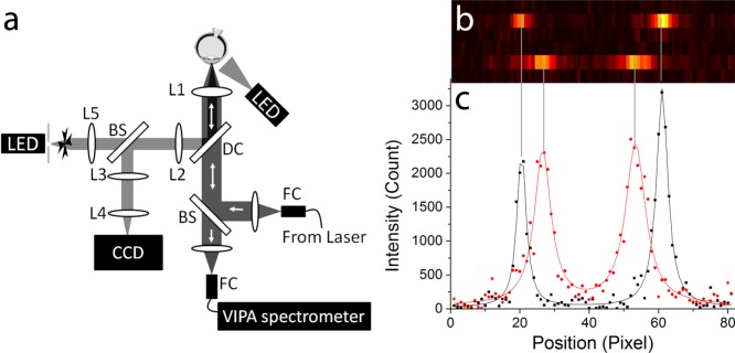
Methods: Brillouin scans were performed along the crystalline lens in 56 eyes from 30 healthy subjects aged from 19 to 63 years. Longitudinal elastic modulus was acquired along the sagittal axis of the lens with a transverse and axial resolution of 4 and 60 μm, respectively. The relative lens stiffness was computed, and correlations with age were analyzed.

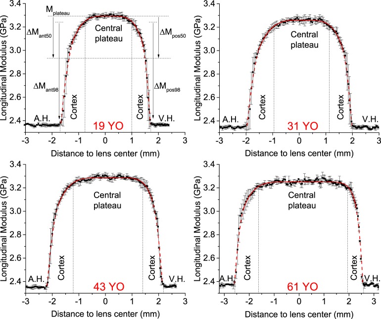
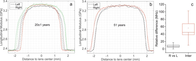
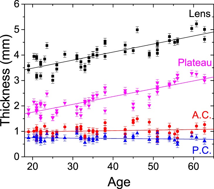
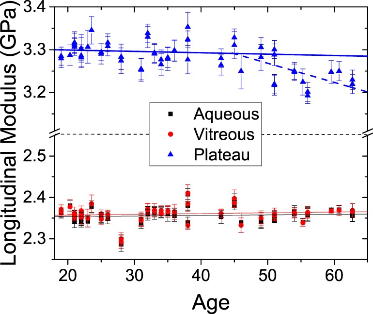
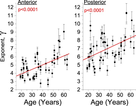
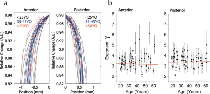
Results: Brillouin axial profiles revealed nonuniform longitudinal modulus within the lens, increasing from a softer periphery toward a stiffer central plateau at all ages. The longitudinal modulus at the central plateau showed no age dependence in a range of 19 to 45 years and a slight decrease with age from 45 to 63 years. A significant intersubject variability was observed in an age-matched analysis. Importantly, the extent of the central stiff plateau region increased steadily over age from 19 to 63 years. The slope of change in Brillouin modulus in the peripheral regions were nearly age-invariant.

Conclusions: The adult human lens showed no measurable age-related increase in the peak longitudinal modulus. The expansion of the stiff central region of the lens is likely to be the major contributing factor to age-related lens stiffening. Brillouin microscopy may be useful in characterizing the crystalline lens for the optimization of surgical or pharmacological treatments aimed at restoring accommodative power.


Figures

Figure 1
Figure 1
(a) Schematic representation of the Brillouin confocal in vivo microscope. (b) Raw EMCCD output of the VIPA spectrometer showing the vitreous humor (top) and lens nucleus (bottom) at an integration time of 0.2 seconds and a laser power of 2 mW. The two peaks represent the stoke shift and the antistoke shift of the following laser order. (c) 2-Lorentzian peak fit (solid line) of the raw imaging data (points) along the Brillouin spectral line for vitreous humor (black) and lens nucleus (red).
Figure 2
Figure 2
Representative Brillouin sagittal profiles in 19-, 31-, 43-, and 60-year-old human lenses showing the aqueous humor (A.H.), the vitreous humor (V.H.), the lens cortex, and central plateau. The central plateau is defined as the top 98% value in longitudinal modulus. The points and error bars represent the mean and SD of successive scans taken along the sagittal axis. Also shown is the definition of the different parameters of the power fit function.
Figure 3
Figure 3
Intra- and intersubject variability. (a) Brillouin sagittal profiles of the left and right eyes of three individuals in an age range of 21 ± 1. (b) Comparison of the left and right eyes in two 51-year-old subjects. (c) The relative difference in the mean longitudinal modulus observed in the central plateau between the left and right eyes in the same subject and between subjects within an age-window of 3 years (Inter). The box and whisker represent the SE and the SD observed in all subjects.
Figure 4
Figure 4
Variation of the human lens anterior cortex (A.C.), posterior cortex (P.C.), central plateau, and overall lens thickness with age. The error bars represent the instrument's thickness accuracy of 120 μm, which is a conservative assumption equal to twice the optical resolution of the Brillouin microscope and accounting for the accuracy in the two interfaces localization.
Figure 5
Figure 5
Variation of the Brillouin longitudinal modulus with age in aqueous humor, vitreous humor, and lens plateau. The error bars represent the SD within each eye subregion. For the plateau region, linear regressions for age younger than 45 years (solid line) and older than 45 years (dashed line) are shown.
Figure 6
Figure 6
Variation of the power law exponent for the anterior and posterior region within the slowly varying central region of the lens. The error bars represent the 95% confidence bound of the fit coefficients.
Figure 7
Figure 7
(a) Comparison of the cortical sagittal elasticity gradient between young (<25 years old), mid (35–45 years old), and old (>50 years old) groups. (b) Variation of the power law exponent for the cortical anterior and posterior regions. The error bars represent the 95% confidence bound of the fit coefficients.
Figure 8
Figure 8
Spatial variation of the shear and longitudinal elastic moduli of ex vivo human lenses along the equatorial axis. (a) Young lenses (n = 2, 4-mm disks from the left and right eyes of a 26-year-old donor). (b) Old lenses (n = 4–8, 4-mm disks from left and right eyes of three donors at ages of 50, 60, and 62). Solid dots, measured data from each individual sample; open squares, average value of the data within each layer; and error bars, standard variation.

References

    1. Duane A. Studies in monocular and binocular accommodation, with their clinical application. Trans Am Ophthalmol Soc. 1922; 20: 132–157. - PMC - PubMed
    1. Strenk SA,, Semmlow JL,, Strenk LM,, Munoz P,, Gronlund-Jacob J,, DeMarco JK. Age-related changes in human ciliary muscle and lens: a magnetic resonance imaging study. Invest Ophthalmol Vis Sci. 1999; 40: 1162–1169. - PubMed
    1. Hermans EA,, Pouwels PJW,, Dubbelman M,, Kuijer JPA,, van der Heijde RGL,, Heethaar RM. Constant volume of the human lens and decrease in surface area of the capsular bag during accommodation: an MRI and Scheimpflug study. Invest Ophthalmol Vis Sci. 2009; 50: 281–289. - PubMed
    1. von Helmholz HH. Handbuch der Physiologishen Optik. Leipzig: Leopold Voss; 1909.
    1. Ostrin LA,, Glasser A. Edinger-Westphal and pharmacologically stimulated accommodative refractive changes and lens and ciliary process movements in rhesus monkeys. Exp Eye Res. 2007; 84: 302–313. - PMC - PubMed