. 2016 Oct 25;55(42):5938-5946.
doi: 10.1021/acs.biochem.6b00858. Epub 2016 Oct 11.

Protein Kinase C Signaling in Adenoviral Infection

Affiliations

Protein Kinase C Signaling in Adenoviral Infection

Mohammad A Yousuf et al. Biochemistry. .

Abstract

Activation of protein kinase C (PKC), a serine/threonine protein kinase, ubiquitously influences cellular signal transduction and has been shown to play a role in viral entry. In this study, we explored a role for PKC in human adenovirus type 37 infection of primary human corneal fibroblasts, a major target cell for infection. We sought evidence for an interaction between PKC activation and two potential downstream targets: cSrc kinase, shown previously to play a critical role in adenovirus signaling in these cells, and caveolin-1, reported earlier to be important to entry of adenovirus type 37. Infection of fibroblasts increased PKCα phosphorylation and translocation of PKCα from the cytosol to caveolin-1 containing vesicles. Virus-induced phosphorylation of both cSrc and AKT was abolished in cell lysates pretreated with calphostin C, a chemical inhibitor of PKC. Inhibition of PKC also reduced virus associated phosphorylation of caveolin-1, while inhibition of cSrc by the chemical inhibitor PP2 reduced only caveolin-1 phosphorylation, but not PKCα phosphorylation, in lipid rafts. These results suggest a role for PKCα upstream to both cSrc and caveolin-1. Phosphorylated PKCα was found in the same endosomal fractions as phosphorylated cSrc, and PKCα was present to a greater degree in caveolin-1 pull downs from virus infected than mock infected cell lysates. Calphostin C also reduced early viral gene expression, indicating that PKCα activity may be required for viral entry. PKCα plays a central role in adenovirus infection of corneal fibroblasts and regulation of downstream molecules, including the important lipid raft component caveolin-1.


Figures

Figure 1
Figure 1
Adenovirus infection of corneal fibroblasts induces activation and translocation of PKCα. (A) SDS-PAGE followed by Western blot analysis for PKC isoforms in serum starved, primary, human corneal fibroblasts shows the presence of the PKCα but not the θ or ζ isoforms. Twenty micrograms of protein was loaded in each well. Anti-GAPDH antibody was also used as a control. The right-most lane is the molecular weight marker. (B) Representative Western blot analysis for phosphorylated and total PKCα upon adenovirus infection. Mock infection of serum-starved fibroblasts with dialysis buffer (M), purified, endotoxin-free adenovirus type 37 (V) at a multiplicity of infection of 10, for 15 min, or adenovirus type 37 following pretreatment with calphostin C (Cal.C + V; 1 µM for 3 h prior to infection) shows increased phosphorylated PKCα (p-PKCα) relative to total PKCα (t-PKCα), and reduction of p-PKCα when cells were pretreated with Cal.C. Virus-infected cells (V) were pretreated with DMSO for 3 h as a control. (C) Densitometric analysis of three consecutive Western blots as in (B), normalized to mock infected levels shows reduction in p-PKCα when fibroblasts are treated with Cal.C prior to infection (*p < 0.05). (D) Immunoconfocal microscopy after infection shows movement of p-PKCα (green) from the plasma membrane to the cytosol within 15 min of infection, and further concentration of p-PKCα in intracytoplasmic vesicles (inset) at 30 min post infection. Isotype control (ctl) treated and others were also stained with DAPI to show cell nuclei.
Figure 2
Figure 2
Viral activation of PKCα occurs upstream of cSrc and AKT and impacts viral entry in human corneal fibroblasts. (A) and (B) show representative Western blots; (C) and (D) show densitometric analysis of three blots as in (A) and (B) for phosphorylated cSrc (p-cSrc; aa: Y416) and AKT (p-AKT; aa: T308) in comparison to total cSrc (t-cSrc) and total AKT (t-AKT) after mock infection (M) with virus-free dialysis buffer, infection with purified adenovirus type 37 (V) for 1 h, or in cells pretreated with calphostin C (1 µM) for 3 h prior to 1 h infection (Cal.C+V). All cells were serum starved and virus-infected cells (V) were pretreated with DMSO for 3 h as a control. The lower blots for t-cSrc and t-AKT were generated by stripping and reprobing the blots above them. Calphostin C pretreatment reduced phosphorylation of both cSrc and AKT otherwise increased by viral infection (*p < 0.05), consistent with an upstream role for PKCα in their activation. (E) Confocal microscopy and (F) quantitative analysis using ImageJ, of viral entry into cells at 30 min post infection, shows reduced entry in cells pretreated with calphostin C (*p < 0.05). Cy3-labeled virus appears red. Cellular actin is stained with phalloidin (green).
Figure 3
Figure 3
Virus infection induces physical association of phosphorylated PKCα (p-PKCα) with caveolin-1 (Cav-1) in whole corneal fibroblast lysates by immunoprecipitation assays (A) and (B), and confocal microscopy (C). Immunoprecipitation (IP) with anticaveolin-1 (A), or anti-p-PKCα (B) followed by immunoblotting (IB) shows increased p-PKCα in caveolin-1 immunoprecipitates and increased caveolin-1 in p-PKCα immunoprecipitates, respectively, at 30 min after viral infection (V), as compared to mock infection (M) with virus-free dialysis buffer. Antirabbit and antimouse IgG isotype controls did not immunoprecipitate either protein. Immunoconfocal microscopy (C) at 30 min post infection demonstrates mostly membrane bound p-PKCα (Alexa Fluor 488: green) and caveolin-1 (Cav-1, Alexa Fluor 568: red) in mock infected cells (M). Virus infected cells (V) show intracellular colocalization of p-PKCα and caveolin-1 in intracytoplasmic vesicles. Cell nuclei are stained with DAPI (blue). Original magnification: 63×.
Figure 4
Figure 4
Inhibition of PKCα activity reduces early viral gene expression of adenovirus type 37 in human corneal fibroblasts. (A) Reverse transcriptase PCR at 4 h after mock (M) or viral (V) infection, with either DMSO or calphostin C (Cal.C+V) pretreatment at 1 µM for 3 h prior to infection. Total RNA was isolated, reverse transcribed, and subjected to PCR with primers specific for E1A and the control gene GAPDH and run on a 1% agarose gel. GAPDH expression is shown, along with reverse transcriptase negative (RT-) controls. E1A expression in virus infected cells is blocked by calphostin C. (B) Representative Western blot (left) and densitometric analysis (right) of three consecutive Western blots show PKCα-specific siRNA knockdown of PKCα protein expression in comparison to untreated (Naïve) cells, and those treated with scRNA (*p < 0.05). (C) Quantitative real-time PCR analysis of E1A expression at 4 h post infection shows almost complete reduction in E1A mRNA expression in cells pretreated with PKCα-specific siRNA (*p < 0.05).
Figure 5
Figure 5
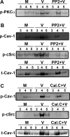
Lipid raft preparations demonstrating increased the presence of p-PKCα, phosphorylated and total caveolin-1 (p-Cav-1 and t-Cav-1, respectively) and phosphorylated cSrc (p-cSrc) in the same raft fractions 1 h after viral infection (V) as compared to mock infection (M), and the effect of chemical inhibition of cSrc by PP2 (A) and (B), and of PKCα by calphostin C (Cal.C) in (C). (A) Western blot for p-PKCα shows increased signal upon viral infection, with slight reduction upon PP2 treatment (10 µM, or DMSO control). (B) Western blot for p-Cav-1, with or without pretreatment by PP2 (or DMSO control), stripped and reprobed for p-cSrc and then stripped and reprobed for t-Cav-1, shows increased p-Cav-1 and p-cSrc in the same lipid raft fractions with a reduction in phosphorylation for both in cells pretreated with PP2. t-Cav-1 in the lipid raft fractions was also slightly reduced by PP2 pretreatment. (C) Western blots on cells pretreated with either DMSO control or calphostin C (1 µM) for 3 h prior to mock or virus infection, and stripped and reprobed as in (B), show a dramatic reduction in p-Cav-1 but not t-Cav-1, and complete abrogation of p-cSrc expression by calphostin C in the same lipid raft fractions.
Figure 6
Figure 6
Activation of cSrc and caveolin-1 in endosomes occurs downstream of PKCα. (A) Western blot analysis of endosomal preparations of serum starved corneal fibroblasts mock infected (M), DMSO (V), or calphostin C pretreated and virus infected (Cal.C+V) for two hr. Postnuclear extracts were subjected to endosome purification using sucrose gradient centrifugation. After SDS-PAGE, membranes were blotted with antibody to p-PKCα, then stripped and reprobed with antibody against phosphorylated cSrc (p-cSrc), and then stripped and reprobed with antibody to phosphorylated caveolin-1 (p-Cav-1). Virus infection increased expression of all three proteins, and all were markedly reduced by pretreatment with calphostin C. (B) cSrc kinase assay performed at 2 h post infection on pooled endosomal preparation fractions 7 through 10 shows increased cSrc activity in virus infection (V) than in mock infection (M) or in virus infected cells pretreated with calphostin C (Cal.C + V, *p < 0.05). Uninfected cells treated with only calphostin C are shown as a control. Note that in this assay, increased kinase activity manifests by reduced fluorescence. (C) Schematic of PKCα signaling initiated by adenovirus infection of corneal fibroblasts.

References

    1. Igumenova TI. Dynamics and Membrane Interactions of Protein Kinase C. Biochemistry. 2015;54:4953–4968. - PMC - PubMed
    1. Constantinescu SN, Cernescu CD, Popescu LM. Effects of protein kinase C inhibitors on viral entry and infectivity. FEBS Lett. 1991;292:31–33. - PubMed
    1. Sieczkarski SB, Brown HA, Whittaker GR. Role of protein kinase C betaII in influenza virus entry via late endosomes. J. Virol. 2003;77:460–469. - PMC - PubMed
    1. San-Juan-Vergara H, Peeples ME, Lockey RF, Mohapatra SS. Protein kinase C-alpha activity is required for respiratory syncytial virus fusion to human bronchial epithelial cells. J. Virol. 2004;78:13717–13726. - PMC - PubMed
    1. Mineo C, Anderson RG. Potocytosis. Robert Feulgen Lecture. Histochem. Cell Biol. 2001;116:109–118. - PubMed

LinkOut - more resources