

Review
doi: 10.1016/j.sbi.2016.09.007.
Epub 2016 Oct 1.
MicroED opens a new era for biological structure determination
Affiliations
- PMID: 27701014
- PMCID: PMC5656569
- DOI: 10.1016/j.sbi.2016.09.007
Item in Clipboard
Review
MicroED opens a new era for biological structure determination
Curr Opin Struct Biol.
2016 Oct.
Abstract
In 2013 we unveiled the cryo-electron microscopy (CryoEM) method of MicroED, or three-dimensional (3D) electron diffraction of microscopic crystals. Here tiny 3D crystals of biological material are used in an electron microscope for diffraction data collection under cryogenic conditions. The data is indexed, integrated, merged and scaled using standard X-ray crystallography software to determine structures at atomic resolution. In this review we provide an overview of the MicroED method and compare it with other CryoEM methods.
Copyright © 2016 Elsevier Ltd. All rights reserved.
Figures
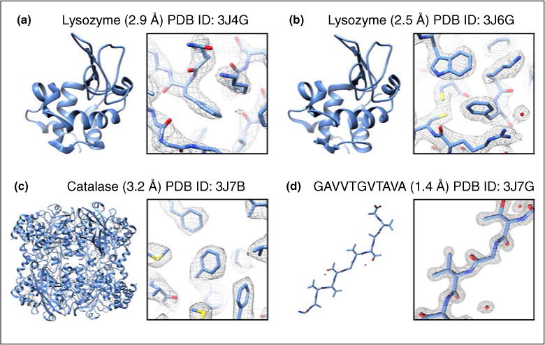
Example structures determined from MicroED. Structures are shown along with their PDB ID, resolution, full model (left) and representative region of the model and density map (right).
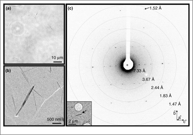
Crystals from the toxic core of α-synuclein. The crystals used to determine the α-synuclein structure were much too small to be seen by light microscopy (a). However, when visualized within the TEM many extremely small microcrystals could be seen (b), which diffracted to approximately 1.4 Å (c). Source: Adapted from Ref. [46••].
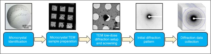
Flow diagram for MicroED data collection protocol. Source: Adapted from Ref. [47•].
References
-
- Zhang YB, Su J, Furukawa H, Yun YF, Gandara F, Duong A, Zou XD, Yaghi OM. Single-crystal structure of a covalent organic framework. J Am Chem Soc. 2013;135:16336–16339. - PubMed
-
- Mugnaioli E, Gorelik T, Kolb U. “Ab initio” structure solution from electron diffraction data obtained by a combination of automated diffraction tomography and precession technique. Ultramicroscopy. 2009;109:758–765. - PubMed
-
- Jiang JX, Jorda JL, Yu JH, Baumes LA, Mugnaioli E, Diaz-Cabanas MJ, Kolb U, Corma A. Synthesis and structure determination of the hierarchical meso-microporous zeolite ITQ-43. Science. 2011;333:1131–1134. - PubMed
Publication types
MeSH terms
Substances
Grants and funding
LinkOut - more resources
Full Text Sources
Other Literature Sources
