. 2016 Nov 16;44(20):9565-9577.
doi: 10.1093/nar/gkw890. Epub 2016 Oct 3.

Laboratory evolution of artificially expanded DNA gives redesignable aptamers that target the toxic form of anthrax protective antigen

Affiliations

Laboratory evolution of artificially expanded DNA gives redesignable aptamers that target the toxic form of anthrax protective antigen

Elisa Biondi et al. Nucleic Acids Res. .

Abstract

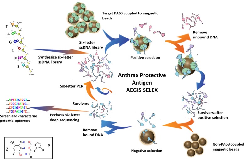
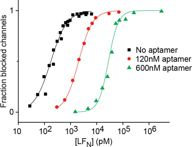
Reported here is a laboratory in vitro evolution (LIVE) experiment based on an artificially expanded genetic information system (AEGIS). This experiment delivers the first example of an AEGIS aptamer that binds to an isolated protein target, the first whose structural contact with its target has been outlined and the first to inhibit biologically important activities of its target, the protective antigen from Bacillus anthracis We show how rational design based on secondary structure predictions can also direct the use of AEGIS to improve the stability and binding of the aptamer to its target. The final aptamer has a dissociation constant of ∼35 nM. These results illustrate the value of AEGIS-LIVE for those seeking to obtain receptors and ligands without the complexities of medicinal chemistry, and also challenge the biophysical community to develop new tools to analyze the spectroscopic signatures of new DNA folds that will emerge in synthetic genetic systems replacing standard DNA and RNA as platforms for LIVE.


Figures

Figure 1.
Figure 1.
Schematic representation of Anthrax Protective Antigen AEGIS SELEX strategy. See main text for description of selection steps. Bottom left insert: molecular structures and hydrogen-bonding pattern for the Z:P pair.
Figure 2.
Figure 2.
PA-Apt1 2D predicted structure and binding to PA. (Left) Percentage of aptamer bound versus PA63 (upper graph) or PA83 (lower graph) concentration obtained in by filter binding assays. The inset in PA63 plot shows calculated binding parameters. Binding parameters for PA83 could not be calculated because signals were below detection level. Plotted are the mean values of three filter-binding assay replicas. (Right) The predicted secondary structure of the PA-Apt1 calculated with KineFold. Nucleotides in the primer binding regions are in light gray circles.
Figure 3.
Figure 3.
Plots of normalized filter binding assay results for all aptamers except PA1T4StdA and PA1T4StdG, for which binding was below the detection level. PA1 (top two graphs) and PA1 + PA1T4 (bottom two graphs) are always present in the experiment, where they serve as a reference for binding of the original aptamer. Plotted are the mean values of three filter-binding assay replicas.
Figure 4.
Figure 4.
DNase I footprinting of PA-Apt1 (PA1). Left: denaturing PAGE of PA-Apt1 DNase I digestions in the presence of increasing concentrations of PA63. First lane: 10-bp DNA ladder with nucleotide numbering on the left. NR1: native PA1, non-reacted. NR2: folded PA1, non-reacted. Position of full-length PA-Apt1 is indicated on the left. Right: PA63-protected nucleotides mapped on the predicted 2D structure of PA1. Green dots: nts of the highly structured ‘core’ aptamer, DNase I-resistant regardless of the presence of PA63. Red dots: positions on the molecule that become protected upon binding to PA63. The 3′ protection boundary is to be regarded as +/− 1 nts due to the lower resolution of the bands on this side of the gel.
Figure 5.
Figure 5.
PA-aptamer variants’ CD signals are indicative of highly structured foldings. Circular dichroism spectra for aptamers PA1, T1 through T6 and PA1 standards tested before (black line) and after (red line) addition of PBS with high Na+ and K+ concentrations.
Figure 6.
Figure 6.
PA1T4-PPZZ optimized AEGIS aptamer against Anthrax protective antigen (PA) PA63. Left, upper: predicted 2D structure and location of P's (red) and Z's (green) non-standard nucleotides. Left, lower: binding on PA63 of PA1T4-PPZZ AEGIS-enriched aptamer compared to its AEGIS-selected, truncated parent molecule. PA1T4-PPZZ presents an observed Kdiss of 35 nM for PA63. Right: binding of PA1T4 (upper) and PA1T4PPZZ (lower) to CMG2–PA63 complex, and binding parameters (inserts).
Figure 7.
Figure 7.
Equilibrium binding competition between LFN and PA1T4 aptamer on PA. Ensemble binding of LFN to PA channels at identical pH 7.6 on both sides of the membrane, with ‘cis’ KCl at 100 mM [added KClcis] and ‘trans’ KCl at 0 mM [added KCltrans] in the absence of the aptamer (black squares), and in the presence of the aptamer at 120 nM (red circles) and 600 nM (green triangles). Curves are fit to a Hill cooperativity model.

Similar articles

Cited by

References

    1. Tuerk C., Gold L. Systematic evolution of ligands by exponential enrichment: RNA ligands to bacteriophage T4 DNA polymerase. Science. 1990;249:505–510. - PubMed
    1. Ellington A.D., Szostak J.W. Selection in vitro of single-stranded DNA molecules that fold into specific ligand-binding structures. Nature. 1992;355:850–852. - PubMed
    1. Battersby T.R., Ang D.N., Burgstaller P., Jurczyk S.C., Bowser M.T., Buchanan D.D., Kennedy R.T., Benner S.A. Quantitative analysis of receptors for adenosine nucleotides obtained via in vitro selection from a library incorporating a cationic nucleotide analog. J. Am. Chem. Soc. 1999;121:9781–9789. - PubMed
    1. Jäger S., Rasched G., Kornreich-Leshem H., Engeser M., Thum O., Famulok M. A versatile toolbox for variable DNA functionalization at high density. J. Am. Chem. Soc. 2005;127:15071–15082. - PubMed
    1. Hollenstein M., Hipolito C.J., Lam C.H., Perrin D.M. A DNAzyme with three protein-like functional groups: enhancing catalytic efficiency of M2+-independent RNA cleavage. Chembiochem. 2009;10:1988–1992. - PubMed

MeSH terms

LinkOut - more resources