. 2016 Oct 4;11(10):e0164105.
doi: 10.1371/journal.pone.0164105. eCollection 2016.

MicroRNA-20b (miR-20b) Promotes the Proliferation, Migration, Invasion, and Tumorigenicity in Esophageal Cancer Cells via the Regulation of Phosphatase and Tensin Homologue Expression

Affiliations

MicroRNA-20b (miR-20b) Promotes the Proliferation, Migration, Invasion, and Tumorigenicity in Esophageal Cancer Cells via the Regulation of Phosphatase and Tensin Homologue Expression

Bin Wang et al. PLoS One. .

Retraction in

Abstract

Increasing evidence has indicated that many microRNAs participate in the development and progression of esophageal cancer and gene expression regulation. MicroRNA-20b (miR-20b) has been reported to be aberrantly expressed in various cancers, but its exact role in esophageal cancer cells remains unclear so far. Therefore, we detected the levels of miR-20b in esophageal tumor tissues and their adjacent normal tissues, and various esophageal cancer cell lines by qRT-PCR. We also explored the effects of miR-20b on cell proliferation, migration, invasion and tumorigenicity of esophageal carcinoma cells through transfection with miR-20b mimics or inhibitor to upregulate or downregulate miR-20b expression in the esophageal cancer cells Eca-109 and KYSE-150, respectively. Additionally, the 3'-untranslated region (3'-UTR) of phosphatase and tensin homologue (PTEN) binding with miR-20b was analyzed by dual-luciferase reporter assays. The results indicated that miR-20b expression level in esophageal tumor tissues was significantly increased compared with their neighboring normal tissues, but its expression was inverse with PTEN protein expression. Luciferase assays confirmed that the 3'-UTR of PTEN was a target of miR-20b in esophageal cancer cells. MiR-20b upregulation promoted cell proliferation, migration, invasiveness, and tumor growth, and decreased apoptosis, and reduced PTEN protein level but not mRNA expression in Eca-109 cells. Conversely, downregulation of miR-20b suppressed these processes in KYSE-150 cells, and enhanced PTEN protein expression. These data indicate that miR-20b plays important roles in tumorigenesis of esophageal cancer possibly via regulation of PTEN expression, and it may be a potential therapeutic target for esophageal cancer treatment.


Conflict of interest statement

The authors have declared that no competing interests exist.

Figures

Fig 1
Fig 1. MiR-20b expression is associated with PTEN level.
(A) The expression levels of miR-20b in 38 paired esophageal tumor tissues and their corresponding adjacent normal tissues were determined by qRT-PCR analyses. * P < 0.05. (B) qRT-PCR analyses of miR-20b expression in human normal esophageal cell line Het-1A, and human esophageal carcinoma cells including TE-1, Eca-109, EC9706, KYSE-150, and SKGT-5. The miR-20b expression was normalized by U6 expression that served as the internal control. * P < 0.05, and ** P < 0.01 compared with Het-1A. (C) The levels of PTEN protein between the esophageal tumor tissues (T) and corresponding adjacent normal tissues (N) were detected by Western blotting. (D) Quantitative analysis of PTEN protein expression that was normalized to β-actin. ** P < 0.01 compared with neighboring normal tissues. (E) The levels of PTEN protein in various cells were detected by Western blot analysis. (F) The relative levels of PTEN protein in various cells were normalized to β-actin. The data represent the mean ± SD (n = 3), * P < 0.05 compared with Het-1A.
Fig 2
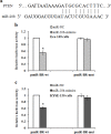
Fig 2. MiR-20b directly targets the 3'-UTR of PTEN.
(A) The position of miR-20b target site in 3'-UTR of PTEN mRNA predicted by miRanda and TargetScan. (B-C) Luciferase assays in Eca-109 cells (B) and KYSE-150 cells (C), and these cells were cotransfected with wt/mut-3'-UTR with miR-20b mimics or negative control (miR-NC). Luciferase activity was detected using dual- luciferase reporter assay system following the manufacturer’s instruction 48 h after transfection. The results were expressed as fold change relative to the negative control. The data represent the mean ± SD (n = 5). * P < 0.05, compared with the corresponding negative control.
Fig 3
Fig 3. MiR-20b inhibits PTEN protein expression and increases p-Akt level.
(A-B) MiR-20b expression was detected by qRT-PCR. A sharp increase of miR-20b expression was observed in the Eca-109 cells transfected with miR-20b mimics (A), while KYSE-150 cells transfected with miR-20b inhibitor resulted in a significant decrease of miR-20b expression (B). (C-D) PTEN mRNA expression was determined by qRT-PCR, and the results indicated that these transfected Eca-109 cells (C) and KYSE-150 cells (D) had no alternation in PTEN mRNA level. (E) The representative images of Western blotting for PTEN, p-Akt and Akt protein expression. (F) Quantitative analysis protein levels of PTEN, p-Akt and Akt by normalization to β-actin. The data represent the mean ± SD (n = 3). * P < 0.05, compared with the corresponding blank control or negative control.
Fig 4
Fig 4. Effects of miR-20b on the proliferation of esophageal carcinoma cells Eca-109 and KYSE-150 cells were transfected with miR-20b mimics and miR-20b inhibitor, respectively.
Cell proliferation was evaluated by MTT assays after the indicated periods. (A) Transfection with miR-20b mimics promoted Eca-109 cell proliferation. (B) Transfection with miR-20b inhibitor suppressed KYSE-150 cell proliferation. The data represent the mean ± SD (n = 3). * P < 0.05, compared with blank control or negative control.
Fig 5
Fig 5. Effects of miR-20b on cellular apoptosis.
Apoptotic percentage in Eca-109 cells or KYSE-150 cells was determined by flow cytometry through the double staining with Annexin V-FITC/ propidium iodide (PI). (A) Representative plots of flow cytometry detection for apoptosis. The percentage of apoptosis was shown in the bottom right quadrant. (B) Apoptotic percentage was quantified and expressed as fold change relative to the corresponding blank control. The data represent the mean ± SD (n = 3). *P < 0.05, #P < 0.05, and P < 0.05.
Fig 6
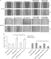
Fig 6. Wound healing assay.
The ability of cell migration was evaluated by wound healing assay after transfection of 48 h. (A) Representative photographs taken at 0 and 24 h after a wound of each group was created by a pipette tip. (B) The number of cells migrated into wound areas was calculated and expressed as fold change relative to the corresponding blank control. The data represent the mean ± SD (n = 3). *P < 0.05, #P < 0.05, and P < 0.05.
Fig 7
Fig 7. Transwell invasion assay.
The ability of cell invasion in vitro was evaluated by Boyden transwell system with porous membranes that were pre-coated with Matrigel after transfection of 48 h. (A) Representative photographs taken at 24 h after the cells were seeded into the upper chamber of transwell, and the cells penetrated through the Matrigel and membranes were fixed in 75% ethanol and stained with hematoxylin and eosin solution. (B) The numbers of cells that penetrated through the membranes to the bottom wells was calculated at five randomly selected visual fields, and expressed as fold change relative to the corresponding blank control. The data represent the mean ± SD (n = 5). * P < 0.05, #P < 0.05, and P < 0.05.
Fig 8
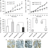
Fig 8. MiR-20 expression promotes tumorigenicity of esophageal carcinoma cells in vivo.
(A-B) Effects of agomir-20b (A) and antagomir-20b (B) on tumor volume in the xenograft model of nude mice. Female BALB/c nude mice were respectively inoculated with Eca-109 cells transfected with agomir-20b or its negative control (agomir-NC), and KYSE-150 cells transfected with antagomir-20b or its negative control (antagomir-NC) on the right flank (n = 5 mice/group). Tumor volume (V) was monitored by measuring the length (L) and width (W) of tumors with a caliper, and calculated with a formula V = π/6 (L × W)3/2. (C) Average weight of tumor tissues. After 40 days, the mice were sacrificed, and the tumor samples were collected and weighted. (D) qRT-PCR analyses of miR-20b expression in tumor tissues. (E) PTEN expression was detected by immunohistochemical staining. The data represent the mean ± SD. * P < 0.05, and ** P < 0.01, agomir-20b vs agomir-NC or antagomir-20b vs antagomir-NC.

References

    1. Li JC, Liu D, Yang Y, Wang XY, Pan DL, Qiu ZD, et al. Growth, clonability, and radiation resistance of esophageal-derived stem-like cells. Asian Pac J Cancer Prev. 2013;14: 4891–4896. - PubMed
    1. Kang M, Li Y, Liu W, Wang R, Tang A, Hao H, et al. miR-129-s suppresses proliferation and migration of esophageal carcinoma cells through downregulation of SOX4 expression. Int J Mol Med. 2013;32: 51–58. 10.3892/ijmm.2013.1384 - DOI - PubMed
    1. Xie YE, Tang EJ, Zhang DR, Ren BX. Down-regulation of Bcl-XL by RNA interference suppresses cell growth and induces apoptosis in humanesophageal cancer cells. World J Gastroenterol. 2006;12: 7472–7477. 10.3748/wjg.v12.i46.7472 - DOI - PMC - PubMed
    1. Zhang HF, Zhang K, Liao LD, Li LY, Du ZP, et al. miR-200b suppresses invasiveness and modulates the cytoskeletal and adhesive machinery in esophageal squamous cell carcinoma cells via targeting Kindlin-2. Carcinogenesis 2014;35: 292–301. 10.1093/carcin/bgt320 - DOI - PubMed
    1. Li D, Ilnytskyy Y, Kovalchuk A, Khachigian LM, Bronson RT, Wang B, et al. Crucial role for early growth response-1 in the transcriptional regulation of miR-20b in breast cancer. Oncotarget 2013;4: 1373–1387. 10.18632/oncotarget.1165 - DOI - PMC - PubMed

Publication types