Review
. 2016 Oct 1;143(19):3434-3448.
doi: 10.1242/dev.139592.

Making sense out of spinal cord somatosensory development

Affiliations
Review

Making sense out of spinal cord somatosensory development

Helen C Lai et al. Development. .

Abstract

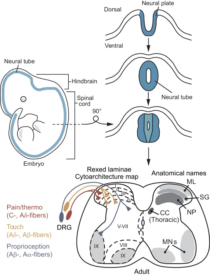
The spinal cord integrates and relays somatosensory input, leading to complex motor responses. Research over the past couple of decades has identified transcription factor networks that function during development to define and instruct the generation of diverse neuronal populations within the spinal cord. A number of studies have now started to connect these developmentally defined populations with their roles in somatosensory circuits. Here, we review our current understanding of how neuronal diversity in the dorsal spinal cord is generated and we discuss the logic underlying how these neurons form the basis of somatosensory circuits.

Keywords: Cutaneous; Dorsal spinal cord development; Itch; Mechanosensation; Neuroepithelium; Nociception; Pain; Proprioception; Pruriception; Thermosensation; Touch; Transcription factor networks; Vertebrate neural tube.


Conflict of interest statement

The authors declare no competing or financial interests.

Figures

Fig. 1.
Fig. 1.
Development of the spinal cord. During development, an invagination of the neural plate closes to form the neural tube, which will become the central nervous system. The most caudal parts of the neural tube will become the spinal cord. Rexed laminae I-X in the adult spinal cord are determined by cytoarchitectonic parameters. Broadly, pain and thermosensitive afferents (C-, Aδ-fibers) from the dorsal root ganglion (DRG) target laminae I-II, touch afferents (Aδ-, Aβ-fibers) target laminae IIinner-V and proprioceptive afferents (Aβ-, Aα-fibers) target more ventral laminae and MNs (see Boxes 1 and 2 for afferent fiber termination markers and definitions). Commonly used anatomical names for Rexed laminae regions are described: marginal layer (ML, lamina I), substantia gelatinosa (SG, lamina II), nucleus proprius (NP, laminae III-V), motor neurons (MNs, lamina IX). Clarke's column, or the dorsal nucleus of Clarke (CC), resides in the medial aspect of lamina VII mainly in the thoracic spinal cord.
Fig. 2.
Fig. 2.
Summary of the transcription factors that set up spinal cord neuronal diversity. The key transcription factors (TFs) that coordinate neuronal diversity in the developing spinal cord are shown, highlighting those that are expressed in the various progenitor domains (dP1-dP6, p0-p3 and pMN) in the proliferating ventricular zone of the developing spinal cord and those that define mature neuronal populations (dI1-6, V0-3 and MN) and their subsets in the differentiating mantle zone. TFs containing a homeodomain are indicated in blue text. Old gene symbols Hb9 (Mnx1), Chx10 (Vsx2), Brn3a (Pou4f1) are shown. Dorsal progenitor (dP), dorsal interneuron 1 contralaterally and ipsilaterally-projecting (dI1c, dI1i), dorsal interneuron late born populations (dILA, dILB), V0 or V3 dorsal, ventral, or cholinergic and glutamatergic (V0D, V0V, V0CG, V3D, V3V), Ia interneuron (IaIN), Vx is an HB9+ population of cells of unknown developmental origin. Msx1, Msx2 (Timmer et al., 2002), Gdf7 (Lee et al., 2000), Atoh1, Neurog1/2, Ascl1, Ptf1a, Pax2, Pax3, Pax6, Pax7, Lbx1, Foxd3, Brn3a, Lhx1/5, Lhx2/9, Barhl1, Barhl2, Isl1, Lmx1b, Phox2a (Bermingham et al., 2001; Ding et al., 2004; Glasgow et al., 2005; Gowan et al., 2001; Gross et al., 2002; Liem et al., 1997; Müller et al., 2002; Saba et al., 2005; Wilson et al., 2008), Dbx1/2, Evx1/2, En1 (Burrill et al., 1997; Moran-Rivard et al., 2001; Pierani et al., 1999, 2001), Olig2/3 (Mizuguchi et al., 2001; Müller et al., 2005; Novitch et al., 2001; Takebayashi et al., 2002), Neurog3 (Sommer et al., 1996), Gsx1/2 (Kriks et al., 2005; Mizuguchi et al., 2006), Lmx1a (Millonig et al., 2000), Nkx6.1/6.2, Nkx2.2/2.9, Irx3, Lhx3, Chx10, Sim1 (Briscoe et al., 2000; Ericson et al., 1997; Fan et al., 1996; Persson et al., 2002), Prdm13 (Chang et al., 2013), Prdm12 (Thelie et al., 2015), Prdm8 (Komai et al., 2009), Gata2/3, Foxn4, Bhlhb5, Pitx2, Foxp1/2, Olig3 (Francius et al., 2013, 2015; Li et al., 2005; Morikawa et al., 2009; Nardelli et al., 1999; Rousso et al., 2008; Skaggs et al., 2011; Zagoraiou et al., 2009), Foxa2 (Ruiz i Altaba et al., 1993), Tlx1/3 (Qian et al., 2002), Prrxl1 (Rebelo et al., 2010), Gbx1 (John et al., 2005), Dmrt3, Wt1 (Andersson et al., 2012; Dyck et al., 2012), Sox1, Sox14, Sox21 (Hargrave et al., 2000; Panayi et al., 2010; Sandberg et al., 2005), Scl (Smith et al., 2002), Hb9, Isl1/2 (Pfaff et al., 1996).
Fig. 3.
Fig. 3.
Dynamic expression of transcription factors in the developing spinal cord. The expression of transcription factors (TFs) in the developing neural tube is highly dynamic. (A) Peak expression of the basic helix-loop-helix (bHLH) transcription factors ATOH1, NEUROG1, ASCL1 and PTF1A within various progenitor domains (dP1-5) in the proliferating ventricular zone occurs at E10.5 and then declines. As these neuronal populations become postmitotic and migrate into the mantle zone, they begin expressing transcription factors that either decline (LHX2/9) or increase (TLX1/3, ISL1, PAX2, LMX1B) over developmental time. It is unknown how FOXD3 expression changes at later development time points (dashed line) (Gross et al., 2002). (B) The interplay between activating bHLH TFs such as ASCL1 and repressive TFs such as HES1, mediated through Notch signaling, results in oscillatory expression of these TFs in neural stem cells; these oscillations control the timing of neurogenesis. Eventually, sustained expression of ASCL1 leads to neuronal differentiation.
Fig. 4.
Fig. 4.
Cross-repression between TFs in the developing neural tube. Morphogens released from the roof plate (BMP, WNT) and floor plate (SHH) set up gradients that impact the expression of TFs in the developing neural tube. For example, dI1-3 (class A) neurons are influenced by BMP signaling, while dI4-6 (class B) neurons are not. Furthermore, cross-repressive activities between individual TFs, both direct and indirect, play an important role in setting up boundaries between interneuron domains.
Fig. 5.
Fig. 5.
Migration of neurons during spinal cord development. Neurons derived from discrete progenitor domains in the developing neural tube migrate quite extensively from their original birth location and do not follow a one-to-one correspondence with Rexed laminae (Fig. 1). For example, while dI1-dI5 neurons remain largely in the dorsal and intermediate spinal cord, dI1-dI3 neurons travel ventrally to the intermediate spinal cord, while dI4/dILA and dI5/dILB neurons migrate dorsally and laterally. V0-V3 populations remain largely ventral, but the V3 domain generates neurons that extend into the dorsal horn. Although dI6 is considered to be dorsally derived, neurons from this domain migrate ventrally (Andersson et al., 2012). Molecular maps represent the current known state of the field.
Fig. 6.
Fig. 6.
Function of neurons arising from dorsal progenitor cells. Neurons derived from a common progenitor source tend to form neurons involved in circuits associated with a particular somatosensory function. Details of these circuits are still under active investigation. (A) Neurons from dI1, dI3 and some of the dI4 domain form networks involved in proprioception, touch-related gross motor and smooth motor control. It is unknown which circuits dI2 lineage neurons produce (dashed line), although some groups suggest they may form SCTs or components of the ALS. By contrast, dI4/dILA and dI5/dILB lineage neurons form circuits involved in pain, temperature, itch and touch. Although dI6 lineage neurons are associated with the developing dorsal neural tube, their known function is in gait motor control in the ventral spinal cord. (B) Summary of the circuits formed by dI1, dI2, dI3, dI4 and dI6 lineage neurons. It is unknown how dI1 and dI2 neurons might project to the medulla, pons, thalamus or other targets of the ALS (?, see text for details). It is also unknown how the axons of dI3 propriospinal neurons travel to the LRt (?, see text for details). (C) Summary of networks formed by dI4/dILA and dI5/dILB neurons. A putative STT in lamina III-VI is of unknown developmental origin (gray circle). Circles outlined in black represent neurons whose soma location is unknown. Excitatory synapses are indicated by solid triangles for monosynaptic connections and open triangles for polysynaptic or unknown monosynaptic connections. Inhibitory synapses are indicated by perpendicular lines at the end of axons. A dashed line indicates the inhibition is indirect. C, contralateral; I, ipsilateral; A, ascending; D, descending; DSCT, VSCT, dorsal/ventral spinocerebellar tract (SCT); ALS, anterolateral system; STT, spinothalamic tract; Prop, proprioceptive; Cut, cutaneous; MN, motor neuron; LRt, lateral reticular nucleus; PSDC, postsynaptic dorsal column; DF, dorsal funiculus; LF, lateral funiculus; VF, ventral funiculus; L, LMX1B+ in lamina I; S, SOM+; R, RORα+; G, GRPR+; V, VGLUT3+; D, DYN+; N, NPY+.

Similar articles

Cited by

References

    1. Abraira V. E. and Ginty D. D. (2013). The sensory neurons of touch. Neuron 79, 618-639. 10.1016/j.neuron.2013.07.051 - DOI - PMC - PubMed
    1. Akay T., Tourtellotte W. G., Arber S. and Jessell T. M. (2014). Degradation of mouse locomotor pattern in the absence of proprioceptive sensory feedback. Proc. Natl. Acad. Sci. USA 111, 16877-16882. 10.1073/pnas.1419045111 - DOI - PMC - PubMed
    1. Alaynick W. A., Jessell T. M. and Pfaff S. L. (2011). SnapShot: spinal cord development. Cell 146, 178-178.e171. 10.1016/j.cell.2011.06.038 - DOI - PMC - PubMed
    1. Alstermark B. and Ekerot C. F. (2013). The lateral reticular nucleus: a precerebellar centre providing the cerebellum with overview and integration of motor functions at systems level. A new hypothesis. J. Physiol. 591, 5453-5458. 10.1113/jphysiol.2013.256669 - DOI - PMC - PubMed
    1. Altman J. and Bayer S. A. (1984). The development of the rat spinal cord. Adv. Anat. Embryol. Cell Biol. 85, 1-166. 10.1007/978-3-642-69537-7_1 - DOI - PubMed

Publication types

Substances