. 2016 Oct 1;143(19):3632-3637.
doi: 10.1242/dev.140137.

Mapping a multiplexed zoo of mRNA expression

Affiliations

Mapping a multiplexed zoo of mRNA expression

Harry M T Choi et al. Development. .

Abstract

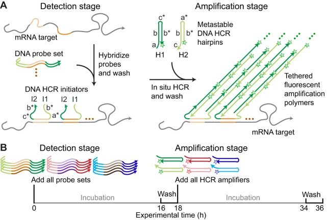
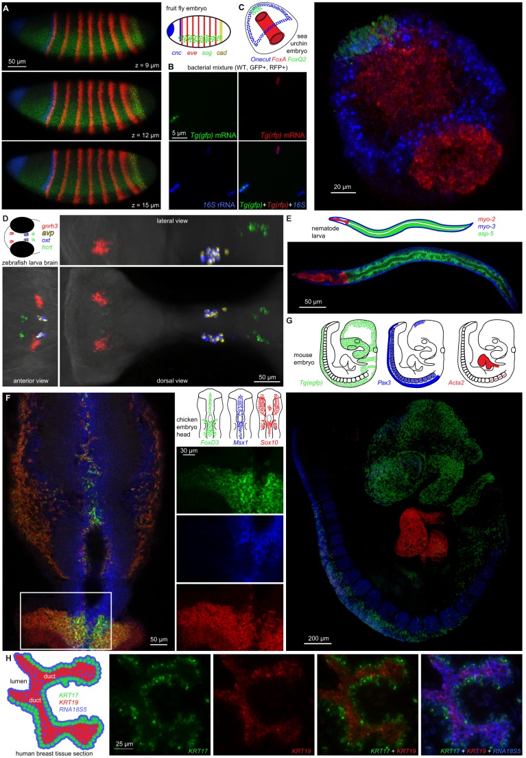
In situ hybridization methods are used across the biological sciences to map mRNA expression within intact specimens. Multiplexed experiments, in which multiple target mRNAs are mapped in a single sample, are essential for studying regulatory interactions, but remain cumbersome in most model organisms. Programmable in situ amplifiers based on the mechanism of hybridization chain reaction (HCR) overcome this longstanding challenge by operating independently within a sample, enabling multiplexed experiments to be performed with an experimental timeline independent of the number of target mRNAs. To assist biologists working across a broad spectrum of organisms, we demonstrate multiplexed in situ HCR in diverse imaging settings: bacteria, whole-mount nematode larvae, whole-mount fruit fly embryos, whole-mount sea urchin embryos, whole-mount zebrafish larvae, whole-mount chicken embryos, whole-mount mouse embryos and formalin-fixed paraffin-embedded human tissue sections. In addition to straightforward multiplexing, in situ HCR enables deep sample penetration, high contrast and subcellular resolution, providing an incisive tool for the study of interlaced and overlapping expression patterns, with implications for research communities across the biological sciences.

Keywords: Bacteria, Whole-mount embryos and larvae; Deep sample penetration; High contrast; Hybridization chain reaction (HCR); In situ amplification; In situ hybridization; Multiplexing; Subcellular resolution; Tissue sections.


Conflict of interest statement

The authors declare competing financial interests in the form of patents and pending patent applications.

Figures

Fig. 1.
Fig. 1.
Multiplexed in situ hybridization chain reaction (HCR). (A) Two-stage in situ HCR protocol (Choi et al., 2014). Detection stage: DNA probes carrying DNA HCR initiators (I1 and I2) hybridize to mRNA targets and unused probes are washed from the sample. Amplification stage: metastable DNA HCR hairpins (H1 and H2) penetrate the sample, initiators trigger chain reactions in which fluorophore-labeled H1 and H2 hairpins sequentially nucleate and open to assemble into tethered fluorescent amplification polymers, and unused hairpins are washed from the sample. See Fig. S1 for a detailed description of the HCR mechanism. (B) Experimental timeline. The time required to perform an experiment is independent of the number of target mRNAs. Stars denote fluorophores.
Fig. 2.
Fig. 2.
Multiplexed mRNA expression maps using in situ HCR. (A) Whole-mount fruit fly (Drosophila melanogaster) embryo: expression schematic and confocal micrographs for four target mRNAs on three planes. Embryo fixed: stage 4-6. (B) Mixed bacterial populations (Escherichia coli: WT, GFP+, RFP+): epifluorescence micrographs (single channels and merge) for three targets (gfp and rfp mRNAs and 16S rRNA). (C) Whole-mount sea urchin embryo (Strongylocentrotus purpuratus): expression schematic and three-dimensional reconstruction from confocal micrographs for three target mRNAs. Embryo fixed: 45 hpf. (D) Whole-mount zebrafish larva (Danio rerio): expression schematic and three-dimensional reconstruction from confocal micrographs for four target mRNAs within the brain. Larva fixed: 5 dpf. (E) Whole-mount nematode larva (Caenorhabditis elegans): expression schematic and confocal micrograph for three target mRNAs. Larva fixed: L3. (F) Whole-mount chicken embryo (Gallus gallus domesticus): expression schematic and confocal micrographs for three target mRNAs in the neural crest (merge and single-channel details). Embryo fixed: stage HH 11-12. (G) Whole-mount mouse embryo [Mus musculus: Tg(Wnt1-Cre; R26R-eGFP)]: expression schematic and three-dimensional reconstruction from confocal micrographs for three target mRNAs. Embryo fixed: E9.5. (H) FFPE human breast tissue section (Homo sapiens sapiens): expression schematic and epifluorescence micrographs for two target mRNAs and one rRNA (single channels and merges). Thickness: 4 µm. See Figs S2-S10 and Movies 1-5 for additional data.
Fig. 3.
Fig. 3.
Subcellular resolution using in situ HCR. (A) Redundant two-channel mapping of target mRNA Acta2 in the heart of a whole-mount mouse embryo. Arrows denote putative sites of active transcription. Probe sets: two probes per channel. Pixel size: 69×69 nm. Embryo fixed: E9.5. (B) Highly correlated intensities for 0.35×0.35 µm voxels in the inset (Pearson correlation coefficient: r=0.92). To avoid inflating the correlation coefficient, we exclude voxels that fall below background thresholds in both channels (excluded voxels lie in the dashed rectangle at the lower left corner of the correlation plot). For each channel, the background threshold is defined as the mean plus two standard deviations for the voxels in the small white square. See Fig. S11 for additional data.

References

    1. Acloque H., Wilkinson D. G. and Nieto M. A. (2008). In situ hybridization analysis of chick embryos in whole-mount and tissue sections. In Avian Embryology, 2nd edn (ed. Bronner-Fraser M.), pp. 169-185. Methods in Cell Biology, vol. 87 San Diego, CA: Elsevier Academic Press. - PubMed
    1. Barroso-Chinea P., Aymerich M. S., Castle M. M., Pérez-Manso M., Tuñón T., Erro E. and Lanciego J. L. (2007). Detection of two different mRNAs in a single section by dual in situ hybridization: A comparison between colorimetric and fluorescent detection. J. Neurosci. Methods 162, 119-128. 10.1016/j.jneumeth.2006.12.017 - DOI - PubMed
    1. Capodieci P., Donovan M., Buchinsky H., Jeffers Y., Cordon-Cardo C., Gerald W., Edelson J., Shenoy S. M. and Singer R. H. (2005). Gene expression profiling in single cells within tissue. Nat. Methods 2, 663-665. 10.1038/nmeth786 - DOI - PMC - PubMed
    1. Chan P. M., Yuen T., Ruf F., Gonzalez-Maeso J. and Sealfon S. C. (2005). Method for multiplex cellular detection of mRNAs using quantum dot fluorescent in situ hybridization. Nucleic Acids Res. 33, e161 10.1093/nar/gni162 - DOI - PMC - PubMed
    1. Chen F., Tillberg P. W. and Boyden E. S. (2015). Expansion microscopy. Science 347, 543-548. 10.1126/science.1260088 - DOI - PMC - PubMed

Publication types