

Significance of TP53 Mutation in Wilms Tumors with Diffuse Anaplasia: A Report from the Children's Oncology Group
- PMID: 27702824
- PMCID: PMC5290091
- DOI: 10.1158/1078-0432.CCR-16-0985
Significance of TP53 Mutation in Wilms Tumors with Diffuse Anaplasia: A Report from the Children's Oncology Group
Abstract
Purpose: To investigate the role and significance of TP53 mutation in diffusely anaplastic Wilms tumors (DAWTs).
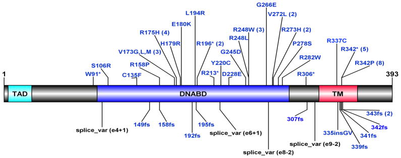
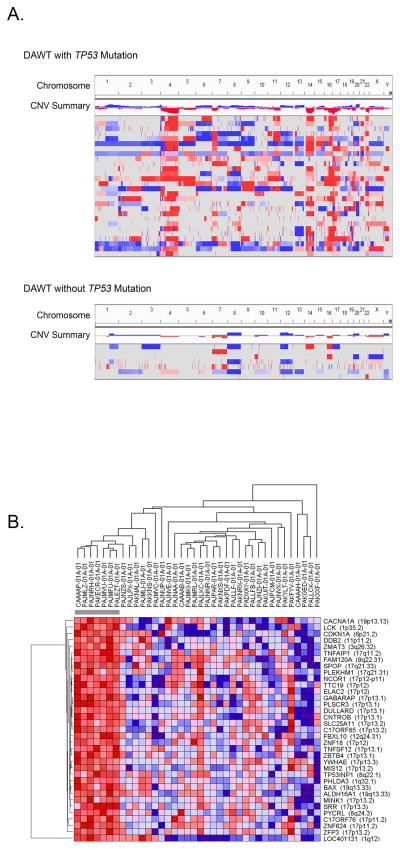
Experimental design: All DAWTs registered on National Wilms Tumor Study-5 (n = 118) with available samples were analyzed for TP53 mutations and copy loss. Integrative genomic analysis was performed on 39 selected DAWTs.
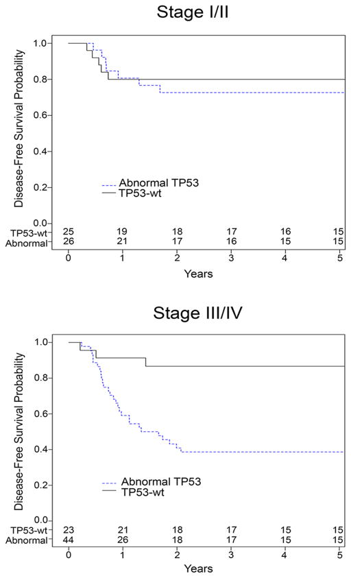
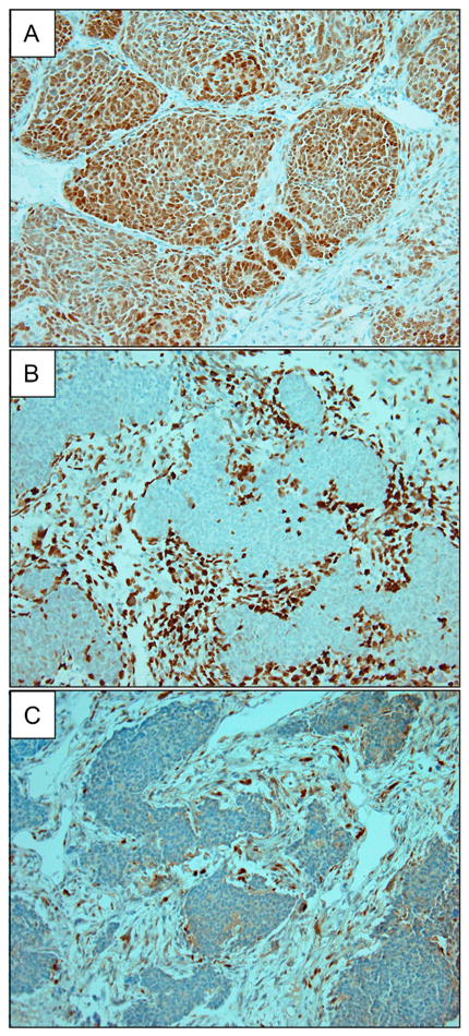
Results: Following analysis of a single random sample, 57 DAWTs (48%) demonstrated TP53 mutations, 13 (11%) copy loss without mutation, and 48 (41%) lacked both [defined as TP53-wild-type (wt)]. Patients with stage III/IV TP53-wt DAWTs (but not those with stage I/II disease) had significantly lower relapse and death rates than those with TP53 abnormalities. In-depth analysis of a subset of 39 DAWTs showed seven (18%) to be TP53-wt: These demonstrated gene expression evidence of an active p53 pathway. Retrospective pathology review of TP53-wt DAWT revealed no or very low volume of anaplasia in six of seven tumors. When samples from TP53-wt tumors known to contain anaplasia histologically were available, abnormal p53 protein accumulation was observed by immunohistochemistry.
Conclusions: These data support the key role of TP53 loss in the development of anaplasia in WT, and support its significant clinical impact in patients with residual anaplastic tumor following surgery. These data also suggest that most DAWTs will show evidence of TP53 mutation when samples selected for the presence of anaplasia are analyzed. This suggests that modifications of the current criteria to also consider volume of anaplasia and documentation of TP53 aberrations may better reflect the risk of relapse and death and enable optimization of therapeutic stratification. Clin Cancer Res; 22(22); 5582-91. ©2016 AACR.
©2016 American Association for Cancer Research.
Conflict of interest statement
The authors disclose no potential conflicts of interest
Figures
References
-
- Howlader NNA, Krapcho M, Garshell J, Miller D, Altekruse SF, et al. In: SEER Cancer Statistics Review, 1975–2012. Cronin KA, editor. National Cancer Institute; 2014. posted to the SEER website, April 2015.
-
- Beckwith JB, Palmer NF. Histopathology and prognosis of Wilms tumors: results from the First National Wilms’ Tumor Study. Cancer. 1978;41(5):1937–48. Epub 1978/05/01. - PubMed
-
- Faria P, Beckwith JB, Mishra K, Zuppan C, Weeks DA, Breslow N, et al. Focal versus diffuse anaplasia in Wilms tumor--new definitions with prognostic significance: a report from the National Wilms Tumor Study Group. The American journal of surgical pathology. 1996;20(8):909–20. Epub 1996/08/01. - PubMed
-
- Dome JS, Cotton CA, Perlman EJ, Breslow NE, Kalapurakal JA, Ritchey ML, et al. Treatment of anaplastic histology Wilms’ tumor: results from the fifth National Wilms’ Tumor Study. Journal of clinical oncology : official journal of the American Society of Clinical Oncology. 2006;24(15):2352–8. doi: 10.1200/jco.2005.04.7852. Epub 2006/05/20. - DOI - PubMed
-
- Pritchard-Jones K, Moroz V, Vujanic G, Powis M, Walker J, Messahel B, et al. Treatment and outcome of Wilms’ tumour patients: an analysis of all cases registered in the UKW3 trial. Annals of oncology : official journal of the European Society for Medical Oncology/ESMO. 2012;23(9):2457–63. doi: 10.1093/annonc/mds025. Epub 2012/03/15. - DOI - PubMed
MeSH terms
Substances
Supplementary concepts
Grants and funding
LinkOut - more resources
Full Text Sources
Other Literature Sources
Medical
Research Materials
Miscellaneous
