. 2017 Jul;37(7):2584-2597.
doi: 10.1177/0271678X16671965. Epub 2016 Jan 1.

Imaging of brain oxygenation with magnetic resonance imaging: A validation with positron emission tomography in the healthy and tumoural brain

Affiliations

Imaging of brain oxygenation with magnetic resonance imaging: A validation with positron emission tomography in the healthy and tumoural brain

Samuel Valable et al. J Cereb Blood Flow Metab. 2017 Jul.

Abstract

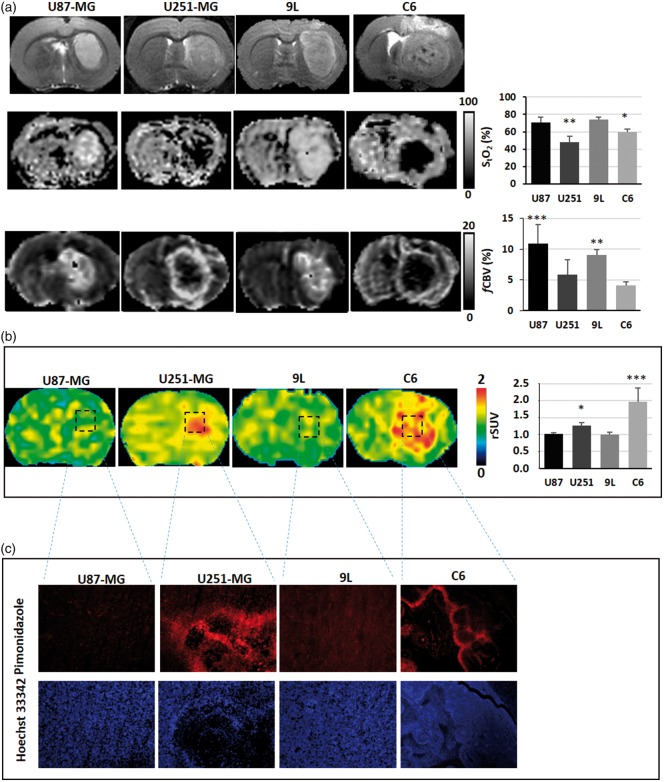
The partial pressure in oxygen remains challenging to map in the brain. Two main strategies exist to obtain surrogate measures of tissue oxygenation: the tissue saturation studied by magnetic resonance imaging (StO2-MRI) and the identification of hypoxia by a positron emission tomography (PET) biomarker with 3-[18F]fluoro-1-(2-nitro-1-imidazolyl)-2-propanol ([18F]-FMISO) as the leading radiopharmaceutical. Nonetheless, a formal validation of StO2-MRI against FMISO-PET has not been performed. The objective of our studies was to compare the two approaches in (a) the normal rat brain when the rats were submitted to hypoxemia; (b) animals implanted with four tumour types differentiated by their oxygenation. Rats were submitted to normoxic and hypoxemic conditions. For the brain tumour experiments, U87-MG, U251-MG, 9L and C6 glioma cells were orthotopically inoculated in rats. For both experiments, StO2-MRI and [18F]-FMISO PET were performed sequentially. Under hypoxemia conditions, StO2-MRI revealed a decrease in oxygen saturation in the brain. Nonetheless, [18F]-FMISO PET, pimonidazole immunohistochemistry and molecular biology were insensitive to hypoxia. Within the context of tumours, StO2-MRI was able to detect hypoxia in the hypoxic models, mimicking [18F]-FMISO PET with high sensitivity/specificity. Altogether, our data clearly support that, in brain pathologies, StO2-MRI could be a robust and specific imaging biomarker to assess hypoxia.

Keywords: Hypoxia; glioblastoma; magnetic resonance imaging; oxygenation; positron emission tomography; rat.


Figures

Figure 1.
Figure 1.
Protocols of the investigations. (A) Assessment of brain oxygenation with various approaches in the healthy brain in normoxic and hypoxemic conditions: (a) protocol with StO2-MRI (n = 6 per condition), (b) protocol with FMISO PET (n = 6 per condition); (B) Assessment of brain oxygenation with multimodal imaging in various tumour models. For each rat, MRI and PET were performed consecutively. Following the MRI examination, the rats were transferred to the PET facilities, intravenously injected with [18F]-FMISO and a PET examination was performed 2 h later. B.G. = blood (arterial) gases.
Figure 2.
Figure 2.
Multiparametric assessment of oxygenation in hypoxemic experiments. (a) Assessment of hypoxia with StO2-MRI (n = 6). Representative T2*-BOLD weighted signal initiated under FiO2 = 30% conditions and continuously monitored when FiO2 was reduced to 17%. Images represent StO2-MRI and fCBV maps of a representative animal during each condition and the graph are the mean ± SD; *p < 0.05 vs FiO2 = 30% using a Student t test. (b) Assessment of hypoxia with [18F]-FMISO PET. Rats were submitted to FiO2 = 30% (n = 6) or FiO2 = 17% (n = 6), [18F]-FMISO was injected and PET images were acquired 2 h later. Results show a representative animal for each condition and the graph are the mean ± SD. (c) Assessment of hypoxia with pimonidazole staining. Representative photographs of pimonidazole staining and the corresponding Hoescht 33342 couterstaining for one animal in each degree of oxygenation.
Figure 3.
Figure 3.
Multiparametric assessment of oxygenation in brain tumour models. (a) Representatives images of rats bearing brain tumours as shown on the T2w MRI (upper panel), and the corresponding assessments of hypoxia with sequential StO2-MRI (middle panel) and fCBV (lower panel) and (b) with [18F]-FMISO PET, or with pimonidazole immunostaining/Hoechst 33342 counterstaining (c). The graphs represent the mean ± SD (n = 6 for U87-MG; n = 7 for 9L; n = 7 for U251-MG and n = 7 for C6); *p < 0.05, **p < 0.01 and ***p < 0.001 vs respective contralateral tissue following a significant ANOVA and Tukey's post-hoc test.
Figure 4.
Figure 4.
Spatial analyses of hypoxia estimates in the C6 glioma model. Spatial inspection of a representative rat for which [18F]-FMISO PET images was segmented so as to obtain a hypoxic volume (upper panel, right); OEF-MRI calculated with StO2-MRI was also segmented (lower panel, right). Pimonidazole mosaic was used as a reference for hypoxia.
Figure 5.
Figure 5.
Comparison of [18F]-FMISO and StO2-MRI. (a) Scatterplot of StO2-MRI values in the tumour ROI of all tumour types as a function of [18F]-FMISO uptake (n = 18 rats who had sequential MRI and PET scans). (b) ROC analyses for both [18F]-FMISO and StO2-MRI using pimonidazole staining as the gold standard.

References

    1. Raichle ME, Gusnard DA. Appraising the brain's energy budget. Proc Natl Acad Sci U S A 2002; 99: 10237–10239. - PMC - PubMed
    1. Baron JC, Marchal G. Ischemic core and penumbra in human stroke. Stroke J Cereb Circ 1999; 30: 1150–1153. - PubMed
    1. Bouzat P, Millet A, Boue Y, et al. Changes in brain tissue oxygenation after treatment of diffuse traumatic brain injury by erythropoietin. Crit Care Med 2013; 41: 1316–1324. - PubMed
    1. Evans SM, Jenkins KW, Chen HI, et al. The relationship among hypoxia, proliferation, and outcome in patients with de novo glioblastoma: A pilot study. Transl Oncol 2010; 3: 160–169. - PMC - PubMed
    1. Bouvier J, Detante O, Tahon F, et al. Reduced CMRO2 and cerebrovascular reserve in patients with severe intracranial arterial stenosis: a combined multiparametric qBOLD oxygenation and BOLD fMRI study. Hum Brain Mapp 2015; 36: 695–706. - PMC - PubMed

MeSH terms