. 2016 Oct 18;113(42):E6535-E6544.
doi: 10.1073/pnas.1611073113. Epub 2016 Oct 4.

Bayesian model reveals latent atrophy factors with dissociable cognitive trajectories in Alzheimer's disease

Collaborators, Affiliations

Bayesian model reveals latent atrophy factors with dissociable cognitive trajectories in Alzheimer's disease

Xiuming Zhang et al. Proc Natl Acad Sci U S A. .

Abstract

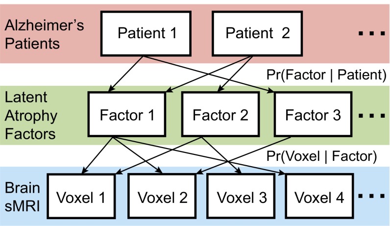
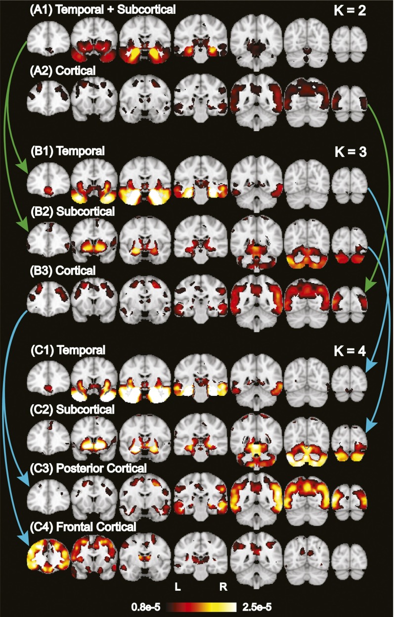
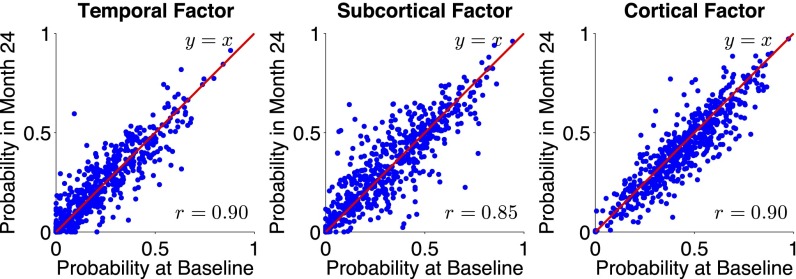
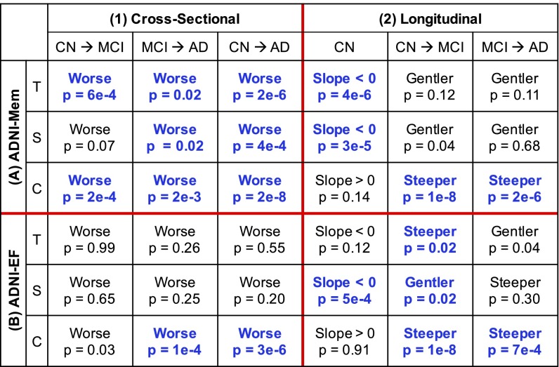
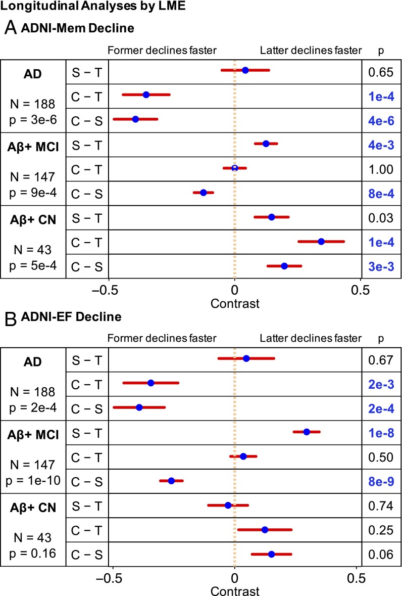
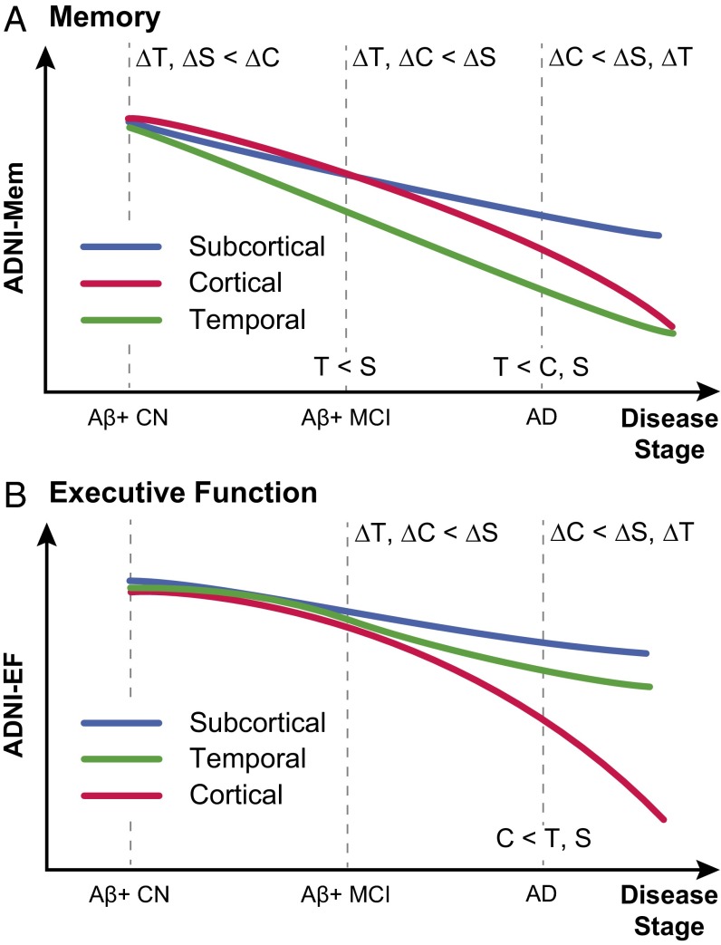
We used a data-driven Bayesian model to automatically identify distinct latent factors of overlapping atrophy patterns from voxelwise structural MRIs of late-onset Alzheimer's disease (AD) dementia patients. Our approach estimated the extent to which multiple distinct atrophy patterns were expressed within each participant rather than assuming that each participant expressed a single atrophy factor. The model revealed a temporal atrophy factor (medial temporal cortex, hippocampus, and amygdala), a subcortical atrophy factor (striatum, thalamus, and cerebellum), and a cortical atrophy factor (frontal, parietal, lateral temporal, and lateral occipital cortices). To explore the influence of each factor in early AD, atrophy factor compositions were inferred in beta-amyloid-positive (Aβ+) mild cognitively impaired (MCI) and cognitively normal (CN) participants. All three factors were associated with memory decline across the entire clinical spectrum, whereas the cortical factor was associated with executive function decline in Aβ+ MCI participants and AD dementia patients. Direct comparison between factors revealed that the temporal factor showed the strongest association with memory, whereas the cortical factor showed the strongest association with executive function. The subcortical factor was associated with the slowest decline for both memory and executive function compared with temporal and cortical factors. These results suggest that distinct patterns of atrophy influence decline across different cognitive domains. Quantification of this heterogeneity may enable the computation of individual-level predictions relevant for disease monitoring and customized therapies. Factor compositions of participants and code used in this article are publicly available for future research.

Keywords: Alzheimer’s disease heterogeneity; Alzheimer’s disease subtypes; mental disorder subtypes; unsupervised machine learning; voxel-based morphometry.


Conflict of interest statement

The authors declare no conflict of interest.

Figures

Fig. 1.
Fig. 1.
A Bayesian model of AD dementia patients, latent atrophy factors, and brain structural MRI (sMRI). Underpinning our approach is the premise that each participant expresses one or more latent factors. Each factor is associated with distinct but possibly overlapping patterns of brain atrophy. The framework can be instantiated with a mathematical model (LDA) (10), with parameters that can be estimated from the structural MRI data of AD dementia patients. The estimated parameters are the probability that a patient expresses a particular factor [i.e., Pr(Factor | Patient)] and the probability that a factor is associated with atrophy at an MRI voxel [i.e., Pr(Voxel | Factor)].
Fig. 2.
Fig. 2.
Hierarchy of latent atrophy factors with distinct atrophy patterns in AD. Bright color indicates higher probability of atrophy at that spatial location for a particular atrophy factor [i.e., Pr(Voxel | Factor)]. Each of the (A) two, (B) three, and (C) four factors was associated with a distinct pattern of brain atrophy and named accordingly. A nested hierarchy of atrophy factors was observed, although the model did not mandate such a hierarchy. For example, when going from two to three factors, the temporal+subcortical factor (A1) split into temporal (B1) and subcortical (B2) factors, whereas the cortical factor remained the same (A2 and B3). From three to four factors, the temporal and subcortical factors remained the same (B1 and C1; B2 and C2), whereas the cortical factor (B3) split into posterior cortical (C3) and frontal cortical (C4) factors. This hierarchical phenomenon was quantified for 2–10 factors (SI Appendix, Fig. S2).
Fig. 3.
Fig. 3.
Stability of factor compositions over two years. Each blue dot represents a participant. For each plot, x and y axes represent the probabilities of factor at baseline and two years after baseline, respectively. In the ideal case, where factor probability estimates remain exactly the same after two years, one would expect a y = x linear fit as well as an r = 1 correlation. In our case, the linear fits were close to y = x with r > 0.85 for all three atrophy factors, suggesting that the factor compositions were stable, despite disease progression.
Fig. 4.
Fig. 4.
Factor compositions of 188 AD dementia patients. Each patient corresponds to a dot, with location (in barycentric coordinates) that represents the factor composition. Color indicates amyloid status: red for Aβ+, green for Aβ−, and blue for unknown. Corners of the triangle represent pure factors; closer distance to the respective corner indicates higher probability for the respective factor. Most dots are far from the corners, suggesting that most patients expressed multiple factors.
Fig. 5.
Fig. 5.
Differences by diagnosis and atrophy factor in (1) cross-sectional baseline and (2) longitudinal decline rates of (A) memory and (B) executive function. Comparisons remaining significant after FDR (q = 0.05) control are highlighted in blue. T, S, and C indicate temporal, subcortical, and cortical factors, respectively. For example, the top left cell of A, 1 suggests that Aβ+ MCI participants with high loading on the temporal factor had worse baseline memory than Aβ+ CN participants with high loading on the same factor (P = 6e−4). However, the bottom left cell of B, 2 suggests that Aβ+ CN participants expressing the cortical factor did not exhibit executive function decline (P = 0.91), whereas the bottom right cell of B, 2 suggests that AD dementia patients expressing the cortical factor showed faster executive function decline than Aβ+ MCI participants expressing the same factor (P = 7e−4).
Fig. 6.
Fig. 6.
Comparisons of baseline (A) memory and (B) executive function in AD dementia patients across factors. Comparisons remaining significant after FDR (q = 0.05) control are highlighted in blue. T, S, and C indicate temporal, subcortical, and cortical factors, respectively. Blue dots are estimated differences between “pure atrophy factors,” and red bars show the SEs (Materials and Methods). For example, the top row in A suggests that AD dementia patients expressing the temporal factor had worse baseline memory than AD dementia patients expressing the subcortical factor (P = 3e−6).
Fig. 7.
Fig. 7.
Comparisons of (A) memory and (B) executive function decline rates across factors by clinical group. Comparisons remaining significant after FDR (q = 0.05) control are highlighted in blue. T, S, and C indicate temporal, subcortical, and cortical factors, respectively. Blue dots are estimated differences between pure atrophy factors, and red bars show the SEs (SI Appendix, SI Methods). For example, the second row in A suggests that AD dementia patients expressing the cortical factor showed faster memory decline than patients expressing the temporal factor (P = 1e−4).
Fig. 8.
Fig. 8.
Schematics of distinct (A) memory and (B) executive function trajectories for temporal, subcortical, and cortical atrophy factors. T, S, and C indicate temporal, subcortical, and cortical factors, respectively. Labels on dotted lines indicate cross-sectional differences. For example, T < C, S in A indicates that the temporal factor was associated with the worst baseline memory among AD dementia patients. Labels in the intervals indicate differences in longitudinal decline rates. For example, ΔT, ΔC < ΔS in B indicates that, among Aβ+ MCI participants, the temporal and cortical factors were associated with faster executive function decline than the subcortical factor. The schematics summarize the behavioral results of Figs. 5, 6, and 7 (more discussion is in SI Appendix, SI Results). Within each cognitive domain, the atrophy factors were associated with distinct trajectories across the stages. The trajectories of the cortical and subcortical factors transpose between the two cognitive domains. Divergence in memory trajectories existed even at the asymptomatic stage of the disease (i.e., among Aβ+ CN participants).

References

    1. Salmon DP, Bondi MW. Neuropsychological assessment of dementia. Annu Rev Psychol. 2009;60:257–282. - PMC - PubMed
    1. Lam B, Masellis M, Freedman M, Stuss DT, Black SE. Clinical, imaging, and pathological heterogeneity of the Alzheimer’s disease syndrome. Alzheimers Res Ther. 2013;5(1):1. - PMC - PubMed
    1. Murray ME, et al. Neuropathologically defined subtypes of Alzheimer’s disease with distinct clinical characteristics: A retrospective study. Lancet Neurol. 2011;10(9):785–796. - PMC - PubMed
    1. Ossenkoppele R, et al. Tau PET patterns mirror clinical and neuroanatomical variability in Alzheimer’s disease. Brain. 2016;139(Pt 5):1551–1567. - PMC - PubMed
    1. Schneider JA, Arvanitakis Z, Leurgans SE, Bennett DA. The neuropathology of probable Alzheimer disease and mild cognitive impairment. Ann Neurol. 2009;66(2):200–208. - PMC - PubMed

Publication types