

Review
doi: 10.1136/bcr-2016-214929.
Acute onset blindness: a case of optic neuritis and review of childhood optic neuritis
Affiliations
- PMID: 27702928
- PMCID: PMC5073568
- DOI: 10.1136/bcr-2016-214929
Item in Clipboard
Review
Acute onset blindness: a case of optic neuritis and review of childhood optic neuritis
BMJ Case Rep.
.
Abstract
Optic neuritis (ON) is an acquired disorder of the optic nerve due to inflammation, demyelination or degeneration. We report a child who presented with acute onset bilateral visual loss who, following a diagnosis of ON, was treated and had excellent visual recovery. Paediatric ON is considered to be different clinical entity to adult ON. Although in children ON is usually parainfectious or postinfectious, it can be the first presenting feature of multiple sclerosis or neuromyelitis optica spectrum disease. In this paper, we discuss the literature on treatment of ON and prediction of risk of recurrence.
2016 BMJ Publishing Group Ltd.
Conflict of interest statement
Conflicts of Interest: None declared.
Figures
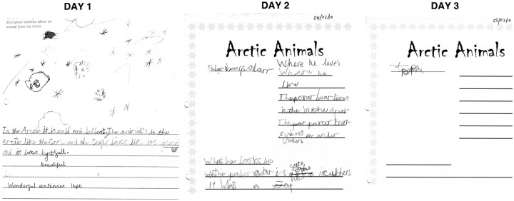
Sample of patient's handwriting showing deterioration over 3 days.
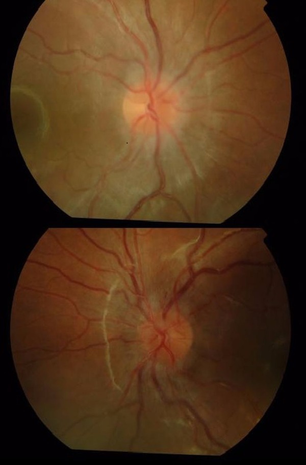
Right and left retinal photographs of posterior pole of the eye, 2 days after presentation. The optic disc is central in the image. There is mild disc swelling and loss of the clear disc margins with softening of the edges.
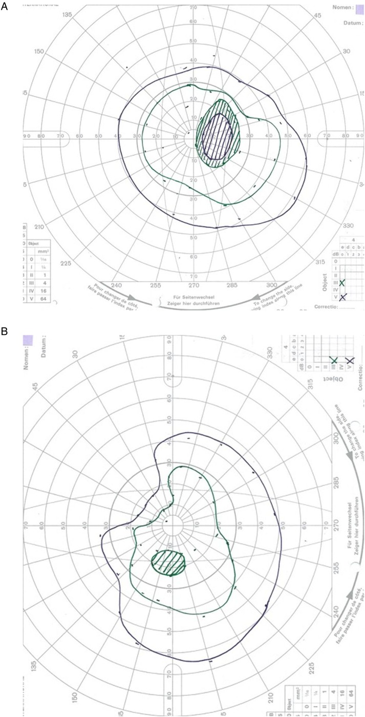
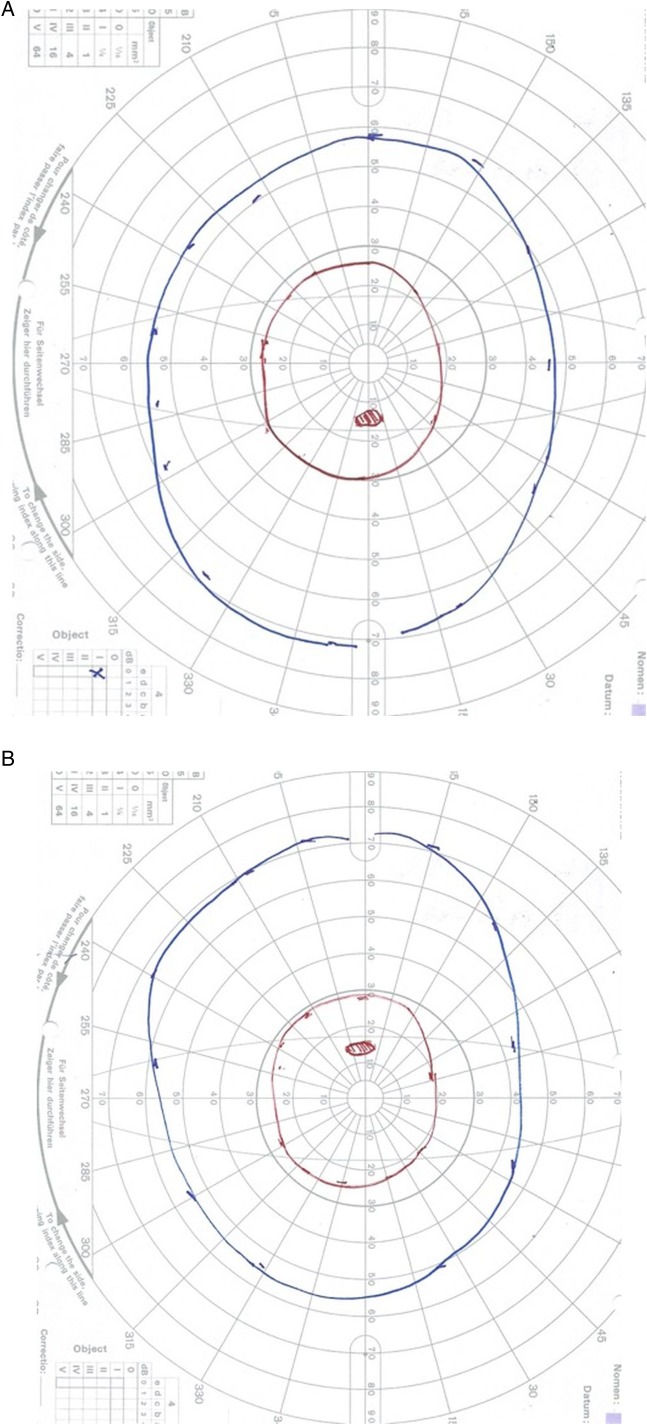
(A and B) Goldmann visual fields in the acute phase showing enlarged blind spot plus constriction in both eyes. The green isopter is III4e, and the purple is V4e.
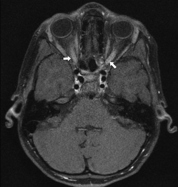
MRI brain with gadolinium showing bilateral postcontrast enhancement of optic nerves.
(A and B) Normal goldmann visual fields in both eyes at 18 months follow-up. The blue isopter is I4e, and the red is I2e. The red is the smaller of the two targets.
Similar articles
-
Longitudinally extensive optic neuritis in neuromyelitis optica spectrum disorder.J Neurol Sci. 2014 Oct 15;345(1-2):209-12. doi: 10.1016/j.jns.2014.07.049. Epub 2014 Jul 28. J Neurol Sci. 2014. PMID: 25125046
-
Clinical features and outcome of childhood optic neuritis at Queen Sirikit National Institute of Child Health.J Med Assoc Thai. 2011 Aug;94 Suppl 3:S189-94. J Med Assoc Thai. 2011. PMID: 22043775
-
Childhood optic neuritis clinical features and outcome.Arch Dis Child. 2011 Sep;96(9):860-2. doi: 10.1136/adc.2009.175422. Epub 2010 Jun 16. Arch Dis Child. 2011. PMID: 20554767
-
Optic neuritis.Compr Ophthalmol Update. 2007 Mar-Apr;8(2):67-75; discussion 77-8. Compr Ophthalmol Update. 2007. PMID: 17540123 Review.
-
[Management of acute visual loss in children].Arch Pediatr. 2004 Nov;11(11):1384-8. doi: 10.1016/j.arcped.2004.04.005. Arch Pediatr. 2004. PMID: 15519841 Review. French.
Cited by
-
Bilateral Pediatric Myelin Oligodendrocyte Glycoprotein Antibody-Associated Disease (MOGAD) Optic Neuritis: A Case Report.Cureus. 2024 Nov 30;16(11):e74883. doi: 10.7759/cureus.74883. eCollection 2024 Nov. Cureus. 2024. PMID: 39741598 Free PMC article.
-
Optic Neuritis Presented as Syndrome of Inappropriate Antidiuretic Hormone Secretion in an 8 Year Old.Case Rep Neurol Med. 2021 Mar 16;2021:6672827. doi: 10.1155/2021/6672827. eCollection 2021. Case Rep Neurol Med. 2021. PMID: 37600468 Free PMC article.
References
-
- Banwell B, Kennedy J, Sadovnick D et al. . Incidence of acquired demyelination of the CNS in Canadian children. Neurology 2009;72:232–9. 10.1212/01.wnl.0000339482.84392.bd - DOI - PubMed
-
- Shams PN, Plant GT. Optic neuritis: a review. Int MS J 2009;16:82–9. - PubMed
-
- Morales DS, Siatkowski RM, Howard CW et al. . Optic neuritis in children. J Pediatr Ophthalmol Strabismus 2000;37:254–9. - PubMed
Publication types
MeSH terms
Substances
LinkOut - more resources
Full Text Sources
Other Literature Sources
Miscellaneous