. 2016 Dec 9;291(50):25965-25982.
doi: 10.1074/jbc.M116.741884. Epub 2016 Oct 4.

Identification of Neutrophil Exocytosis Inhibitors (Nexinhibs), Small Molecule Inhibitors of Neutrophil Exocytosis and Inflammation: DRUGGABILITY OF THE SMALL GTPase Rab27a

Affiliations

Identification of Neutrophil Exocytosis Inhibitors (Nexinhibs), Small Molecule Inhibitors of Neutrophil Exocytosis and Inflammation: DRUGGABILITY OF THE SMALL GTPase Rab27a

Jennifer L Johnson et al. J Biol Chem. .

Abstract

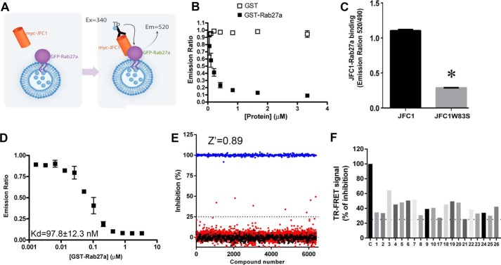
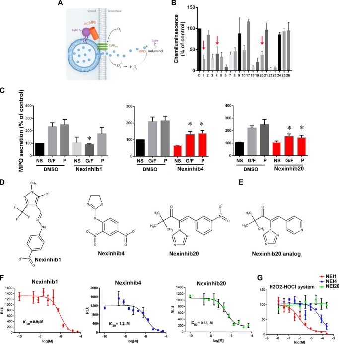
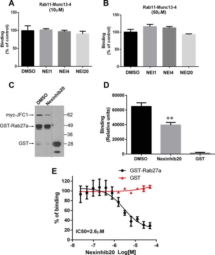
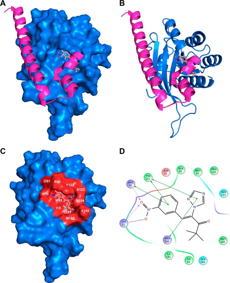
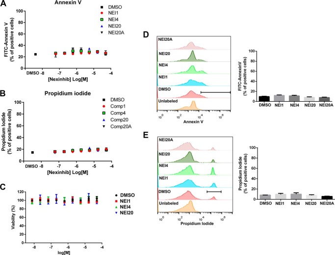
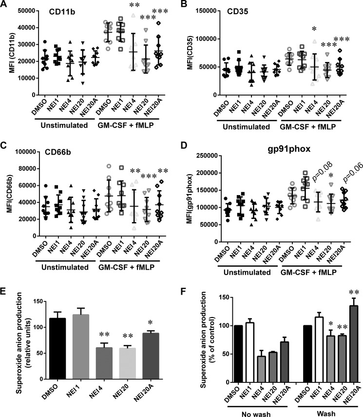
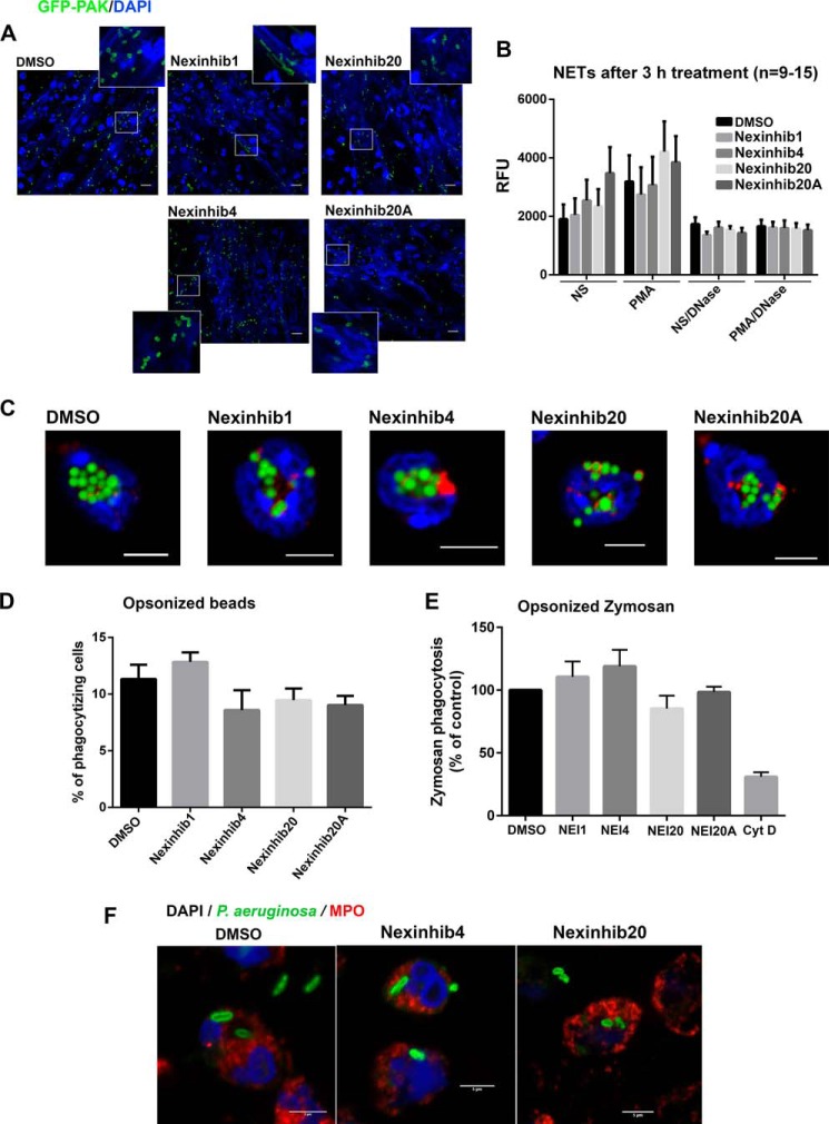
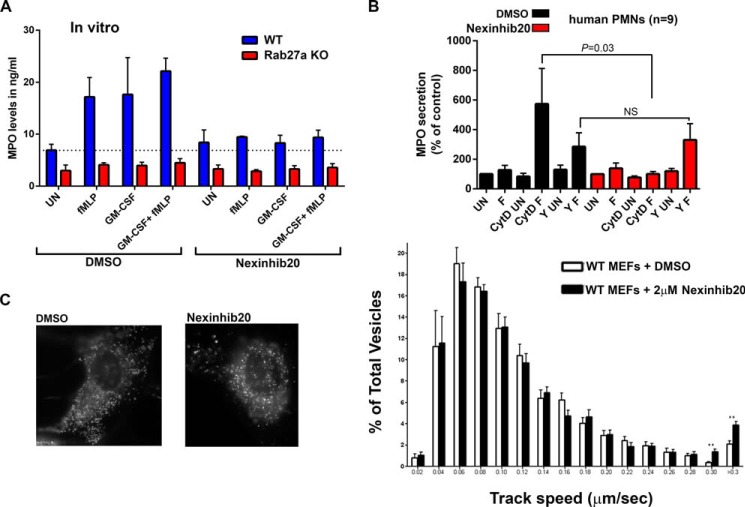
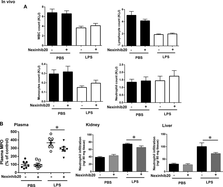
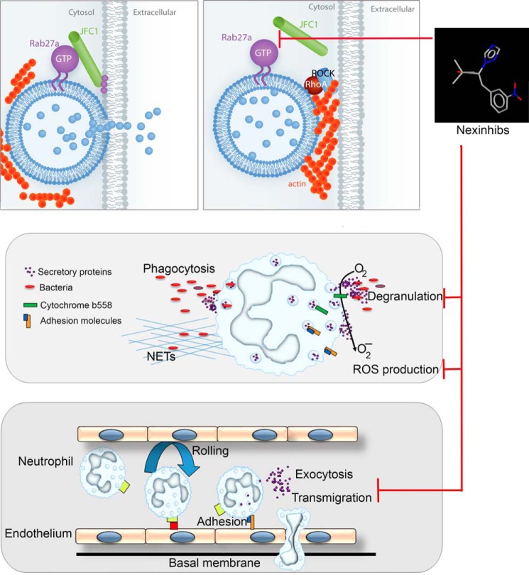
Neutrophils constitute the first line of cellular defense in response to bacterial and fungal infections and rely on granular proteins to kill microorganisms, but uncontrolled secretion of neutrophil cargos is injurious to the host and should be closely regulated. Thus, increased plasma levels of neutrophil secretory proteins, including myeloperoxidase and elastase, are associated with tissue damage and are hallmarks of systemic inflammation. Here, we describe a novel high-throughput screening approach to identify small molecule inhibitors of the interaction between the small GTPase Rab27a and its effector JFC1, two central regulators of neutrophil exocytosis. Using this assay, we have identified small molecule inhibitors of Rab27a-JFC1 binding that were also active in cell-based neutrophil-specific exocytosis assays, demonstrating the druggability of Rab GTPases and their effectors. These compounds, named Nexinhibs (neutrophil exocytosis inhibitors), inhibit exocytosis of azurophilic granules in human neutrophils without affecting other important innate immune responses, including phagocytosis and neutrophil extracellular trap production. Furthermore, the compounds are reversible and potent inhibitors of the extracellular production of superoxide anion by preventing the up-regulation of the granule membrane-associated subunit of the NADPH oxidase at the plasma membrane. Nexinhibs also inhibit the up-regulation of activation signature molecules, including the adhesion molecules CD11b and CD66b. Importantly, by using a mouse model of endotoxin-induced systemic inflammation, we show that these inhibitors have significant activity in vivo manifested by decreased plasma levels of neutrophil secretory proteins and significantly decreased tissue infiltration by inflammatory neutrophils. Altogether, our data present the first neutrophil exocytosis-specific inhibitor with in vivo anti-inflammatory activity, supporting its potential use as an inhibitor of systemic inflammation.

Keywords: JFC1; Rab27a GTPases and effectors; druggability of GTPases; exocytosis; inflammation; inhibitor; innate immunity; lipopolysaccharide (LPS); neutrophil; systemic inflammation.


Figures

FIGURE 1.
FIGURE 1.
High-throughput screening for the identification of inhibitors of the JFC1-Rab27a interaction. A, schematic representation of the TR-FRET binding reaction. Cell lysates expressing Myc-JFC1 or EGFP-Rab27a were mixed and incubated with terbium-conjugated anti-Myc antibody. The samples were excited at 340 nm. The emission peak of terbium (centered at 490 nm) overlaps with the excitation spectrum of GFP. FRET signal was measured by detecting GFP emission at 520 nm, and results are expressed as the emission ratio of the acceptor (GFP, 520 nm)/donor (terbium, 490 nm, used as internal control). An increased emission ratio is indicative of specific binding. B, specific signal of the Myc-JFC1/EGFP-Rab27a TR-FRET reaction was inhibited by recombinant-purified GST-Rab27a but not GST. The baseline reading for the reaction in the absence of GST or GST-Rab27a was 1.050 ± 0.005. C, single amino acid mutant JFC1-W83S has decreased signal in the TR-FRET assay. Mean ± S.E. from triplicates of one experiment representative of three experiments. *, p < 0.001. D, homologous competitive binding experiments for Rab27a using the TR-FRET assay. Specific binding of a constant concentration of EGFP-Rab27a in the presence of various concentrations of GST-Rab27a was measured. IC50 values were determined using appropriate concentrations (12.5, 25, or 50 nm) of EGFP-Rab27a, so the concentration of EGFP-Rab27a was less than half the IC50. Kd value was then calculated using the homologous competitive binding curve fitted to a built-in equation of one-site competition (GraphPad Prism). The assay assumes that GST-Rab27a and EGFP-Rab27a have similar affinity for JFC1. Error bars correspond to S.E. of three replicates. E, HTS for small molecule inhibitors of the JFC1-Rab27a interaction were performed using the Maybridge HitFinder library. Compounds (red circles) or DMSO (black circles) were added by pin tool into 384-well plates containing lysates expressing Myc-JFC1 and incubated for 15 min at 20 °C. Next, EGFP-Rab27a or EGFP (negative control, blue circles) expressing lysates were loaded using a liquid handling device, and samples were further incubated for 15 min. Reactions were started by addition of terbium-conjugated anti-Myc antibody, and TR-FRET signal was measured by detecting the ratio of the acceptor (GFP, 520 nm)/donor (terbium, 490 nm). Compounds found to inhibit binding exceeding the 3-σ statistical limit (dotted line) were considered primary positive hits. F, inhibitory activity of 20 compounds chosen for follow-up experiments. Compounds 1, 4, and 20 correspond to Nexinhib1, -4, and -20.
FIGURE 2.
FIGURE 2.
Identification of neutrophil exocytosis-specific inhibitors. A, schematic representation of the cell-based secondary screening used for the identification of cell-active inhibitors of MPO secretion. B, 13 compounds, inhibitors in the parent TR-FRET assay, were identified as cell actives in the PMA-induced MPO secretion assay. Mean ± S.E. from three independent experiments using neutrophils from independent donors. The three compounds that were further followed up based on their inhibitory effect on MPO secretion assays (C) are indicated with arrows. C, analysis of MPO secretion. Human neutrophils were treated with the indicated compounds for 1 h at 10 μm and subsequently stimulated with GM-CSF and fMLP (G/F), PMA (P), or left untreated (NS). MPO secretion was analyzed by ELISA. Three inhibitors were identified. Mean ± S.E. from 4 to 11 independent experiments using neutrophil from different healthy donors. *, p < 0.05. D, molecular structures of Nexinhibs. E, molecular structure of Nexinhib20 analog. F, dose-response inhibitory activity of Nexinhibs using the chemiluminescence-based MPO secretion assay. Mean ± S.E. of three biological replicates. G, cell-free luminescence assay showing scavenger activity of Nexinhib (NEI) 1, low scavenger activity of Nexinhib4, and no scavenger activity of Nexinhib20.
FIGURE 3.
FIGURE 3.
Nexinhibs specifically inhibit Rab27a-JFC1 binding but do not interfere with the interaction of other Rab GTPases and effectors. A and B, counterscreening TR-FRET assay for Rab11 and Munc13-4 shows lack of activity for Nexinhibs (NEI) on Rab11-Munc13-4 binding either at 10 μm (A) or at 50 μm (B). C, orthogonal validation pulldown assay showing significant inhibition of Rab27a-JFC1 binding by Nexinhib20 (50 μm). D, quantification of pulldown assay shown in C. Mean ± S.E. from three independent experiments. E, ELISA approach to determine the potency of Nexinhib20 to inhibit the Rab27a-JFC1 interaction performed in dose-response format using recombinant GST-Rab27a or GST (negative control) to bind to JFC1. The experiments were performed in triplicate using a 12-point 2-fold serial dilutions of the hit compounds in DMSO. Mean ± S.E. of three independent experiments.
FIGURE 4.
FIGURE 4.
Molecular docking analysis of Nexinhib20 on Rab27a suggests its mechanism of Rab27a/JFC1 disruption. A, surface representation of Rab27a (blue) in complex with Nexinhib20 shown as sticks in the best scoring pose compared with the binding of Exophilin4 (Slp2a) (magenta, Protein Data Bank code 3BC1). B, schematic representation of RAb27a bound to Nexinhib20 as in A. C, Rab27a binding surface of Nexinhib20 is identified in red. The Rab27a residues interacting with the Nexinhib20 compound are indicated. D, two-dimensional schematic representation of the Rab27a and Nexinhib20 complex interactions. Green lines indicate π-π stacking interactions; purple dashed arrows represent side-chain hydrogen bond interactions. Positively charged, negatively charged, polar, and hydrophobic residues are depicted with blue, red, cyan, and green circles, respectively.
FIGURE 5.
FIGURE 5.
Nexinhibs do not induce apoptosis or cell death. A and B, dose-response analyses of the effect of compounds on apoptosis and cell viability, respectively. Cells were incubated in the presence of Nexinhibs (NEI) at the indicated concentrations for 1 h. A, early signs of apoptosis were established by staining phosphatidylserine at the outer leaflet of the plasma membrane using FITC annexin V after treatment with compounds or DMSO. B, cell death in compound- and DMSO-treated groups was analyzed by incorporation of propidium iodide (PI) as described under “Materials and Methods.” C, metabolic activity was measured in a dose-response format 1 h after treatment of human neutrophils with compounds or DMSO using a luminescent cell viability assay for the quantification of ATP, as described under “Materials and Methods.” D and E, analysis of early apoptosis and cell death was performed as in A and B, respectively, 4 h after treatment with the indicated compounds at 10 μm. Left panels, representative histograms from flow cytometry analyses of annexin V (D) or propidium iodide (E) are shown. Right panels, the number of FITC annexin V-positive (D) or propidium iodide-positive cells (E) after compound or DMSO treatment was quantified, and results were expressed as % of positive cells for each experimental condition. A–E, mean ± S.E., n = 3.
FIGURE 6.
FIGURE 6.
Functional analyses identify Nexinhibs as efficient inhibitors of neutrophil exocytosis and extracellular superoxide anion production. A–D, up-regulation of the neutrophil adhesion molecule CD11b, the receptors CD66b and CD35, and the NADPH oxidase membrane-associated subunit gp91phox at the plasma membrane was evaluated by flow cytometry using specific antibodies that detect extracellular epitopes of the indicated markers. Human neutrophils were incubated with the indicated Nexinhibs (NEI) at 10 μm or DMSO and subsequently treated with GM-CSF and fMLP or vehicle (Unstimulated). Symbols correspond to individual donors, and error bars indicate mean ± S.E. of nine independent donors. *, p < 0.02; **, p < 0.003; ***, p < 0.0003, versus DMSO, stimulated. Paired Student's t test. E, production of extracellular superoxide anion by phorbol ester-stimulated human neutrophils treated either with the indicated compounds at 10 μm or vehicle for 1 h was analyzed using the cytochrome c reduction assay. Mean ± S.E. from 7 to 10 independent donors. *, p < 0.04; **, p < 0.001, versus DMSO. Unpaired Student's t test. F, superoxide anion production was measured as in E except that, where indicated, the Nexinhib or DMSO-treated cells were washed twice in PBS before the addition of stimuli. Mean ± S.E. (n = 4). *, p < 0.02; **, p < 0.002, versus same compound, no wash condition.
FIGURE 7.
FIGURE 7.
Nexinhibs do not interfere with NET production or phagocytosis. A, Nexinhib-treated human neutrophils produce NETs (DNA staining, blue, DAPI) and trap bacteria (P. aeruginosa, red). Scale bar, 10 μm. B, compound-treated neutrophils respond to PMA by producing NETs. Human neutrophils were stimulated with PMA for 3 h in the presence of the indicated compound or vehicle. Where indicated, the reactions were performed in the presence of DNase to dismantle NETs as a negative control. NETs were analyzed using the cell-impermeant DNA-staining probe SYTOX Green and quantified by fluorometry. Mean ± S.E. (n = 9–15). C, confocal microscopy analysis of the phagocytosis of opsonized fluorescent latex beads by mouse neutrophils after treatment with the indicated compounds at 10 μm or vehicle for 1 h. Green, beads; red, MPO; blue, DAPI. Scale bar, 5 μm. D, quantification of phagocytosis by flow cytometry. Experiments were performed as in C. Mean ± S.E. of neutrophil samples from six independent mice. E, phagocytosis of opsonized zymosan by human neutrophils after treatment with the indicated compounds at 10 μm or vehicle for 1 h. The data represent the mean ± S.E. from five independent donors. Cyt D, cytochalasin D. F, confocal microscopy analysis of the phagocytosis of opsonized live bacteria by human neutrophils after treatment with the indicated compounds at 10 μm or vehicle for 1 h. Scale bar, 5 μm.
FIGURE 8.
FIGURE 8.
Mechanism of Nexinhib20 regulation of neutrophil exocytosis. A, Nexinhib20 inhibits the exocytosis of azurophilic granule to basal levels similar to those observed for Rab27a-KO neutrophils. Neutrophils from wild type or Rab27a-KO mice were incubated with 10 μm Nexinhib20 or vehicle for 1 h; the cells were stimulated with the indicated stimuli, and secreted MPO was quantified by ELISA. Mean ± S.E. (n = 3). B, Nexinhibs inhibit MPO secretion upstream of JFC1-dependent inactivation of the RhoA pathway. Human neutrophils were treated with 10 μm Nexinhib20 or vehicle for 1 h. Subsequently, the cells were incubated in the presence of 10 μg/ml cytochalasin D (CytD), with the ROCK kinase inhibitor Y27632 (Y) or DMSO and stimulated with fMLP (F) or left untreated (UN). Mean ± S.E. (n = 9). C, Nexinhib20 interferes with vesicular docking at the plasma membrane. Mouse embryonic fibroblasts were transfected for the expression of the lysosomal marker LysoTracker. Vesicular trafficking in the plane parallel to the plasma membrane (exocytic active zone) was analyzed by TIRFM. Left panel, representative images showing that Nexinhib20 does not alter the overall lysosomal distribution. Right panel, quantitative analysis of vesicular dynamics in DMSO or Nexinhib20-treated cells. Histograms representing the speeds of lysosomes in DMSO- (white bars) or compound (black bars)-treated cells are shown. The speeds for the independent vesicles were binned in 0.02-μm/s increments and plotted as a percentage of total vesicles for a given cell. Results are represented as mean ± S.E. from 40 DMSO- and 40 compound-treated cells. **, p < 0.01. n = 3.
FIGURE 9.
FIGURE 9.
Nexinhib20 has a protective effect in a mouse model of endotoxin-induced systemic inflammation. Mice were injected (i.p.) with Nexinhib20 (30 mg/kg) or vehicle 3 h before insult with a single intraperitoneal dose of LPS (E. coli 0111:B4 Alexis Biochemicals) (7.5 mg/kg), and blood and tissues were collected 4 h post-LPS administration. A, blood cell counts, mean ± S.E. (n = 7). B, effect of Nexinhib20 on LPS-induced systemic neutrophil secretion and tissue infiltration. Left panel, quantification of plasma levels of neutrophil-secreted MPO was performed by ELISA and expressed as percentage of control (PBS). Mean ± S.E. *, p < 0.05 (n = 7). Center and right panels, Nexinhib20 decreases neutrophil infiltration into tissues in a model of endotoxemia. Mice were treated as above, and the level of neutrophil infiltration into the indicated tissues was analyzed by the quantification of total tissue myeloperoxidase. Significant decrease in neutrophil infiltration was observed in kidneys (p < 0.05) and liver (p = 0.05). Mean ± S.E. (n = 7).
FIGURE 10.
FIGURE 10.
Mechanism of action of neutrophil exocytosis inhibitors, Nexinhibs. Upper panels, left, the small GTPase Rab27a interacts with its effector JFC1 to facilitate vesicular trafficking and cargo secretion. Right, Nexinhib20 inhibits the Rab27a-JFC1 binding and impairs exocytosis possibly by an indirect increase in RhoA signaling, a process up-regulated in JFC1-deficient cells (18). Middle, inhibition of neutrophil exocytosis impairs both neutrophil cargo release and the up-regulation of important granule membrane-associated regulatory proteins, including the cytochrome b558 of the NADPH oxidase, with the consequent decrease of extracellular ROS production. Lower panel, in vivo inhibition of neutrophil exocytosis decreases plasma levels of neutrophil toxic proteins during systemic inflammation and decreases transmigration into tissues, most likely by inhibition of the up-regulation of adhesion molecules.

Similar articles

Cited by

References

    1. Segal A. W. (2005) How neutrophils kill microbes. Annu. Rev. Immunol. 23, 197–223 - PMC - PubMed
    1. Mantovani A., Cassatella M. A., Costantini C., and Jaillon S. (2011) Neutrophils in the activation and regulation of innate and adaptive immunity. Nat. Rev. Immunol. 11, 519–531 - PubMed
    1. Lee W. L., Harrison R. E., and Grinstein S. (2003) Phagocytosis by neutrophils. Microbes Infect. 5, 1299–1306 - PubMed
    1. Brinkmann V., Reichard U., Goosmann C., Fauler B., Uhlemann Y., Weiss D. S., Weinrauch Y., and Zychlinsky A. (2004) Neutrophil extracellular traps kill bacteria. Science 303, 1532–1535 - PubMed
    1. Amulic B., Cazalet C., Hayes G. L., Metzler K. D., and Zychlinsky A. (2012) Neutrophil function: from mechanisms to disease. Annu. Rev. Immunol. 30, 459–489 - PubMed

Associated data