. 2016 Oct 5:6:33987.
doi: 10.1038/srep33987.

Epigraph: A Vaccine Design Tool Applied to an HIV Therapeutic Vaccine and a Pan-Filovirus Vaccine

Affiliations

Epigraph: A Vaccine Design Tool Applied to an HIV Therapeutic Vaccine and a Pan-Filovirus Vaccine

James Theiler et al. Sci Rep. .

Abstract

Epigraph is an efficient graph-based algorithm for designing vaccine antigens to optimize potential T-cell epitope (PTE) coverage. Epigraph vaccine antigens are functionally similar to Mosaic vaccines, which have demonstrated effectiveness in preliminary HIV non-human primate studies. In contrast to the Mosaic algorithm, Epigraph is substantially faster, and in restricted cases, provides a mathematically optimal solution. Epigraph furthermore has new features that enable enhanced vaccine design flexibility. These features include the ability to exclude rare epitopes from a design, to optimize population coverage based on inexact epitope matches, and to apply the code to both aligned and unaligned input sequences. Epigraph was developed to provide practical design solutions for two outstanding vaccine problems. The first of these is a personalized approach to a therapeutic T-cell HIV vaccine that would provide antigens with an excellent match to an individual's infecting strain, intended to contain or clear a chronic infection. The second is a pan-filovirus vaccine, with the potential to protect against all known viruses in the Filoviradae family, including ebolaviruses. A web-based interface to run the Epigraph tool suite is available (http://www.hiv.lanl.gov/content/sequence/EPIGRAPH/epigraph.html).


Conflict of interest statement

K.F., L.J.P., B.K. and J.T. are co-inventors on a patent application (PCT/US15/54067) on “HIV Vaccines Comprising One or More Population Episensus Antigens.” Authors LJP and KF have a significant financial interest in TomegaVax Inc., a company that may have a commercial interest in the results of this research and technology. This potential individual and institutional conflict of interest has been reviewed and managed by OHSU.

Figures

Figure 1
Figure 1
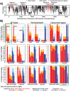
(a)Full graph for the CRF01-AE clade of the Nef protein. The green rectangle is an inset shown in (B). Nodes are red dots, and represent each k-mer variant, with k = 9. The edges are thin blue lines that connect epitopes whose sequences overlap by k − 1 amino acids, as shown for the first two epitopes (ea = VTSSNMNNA, eb = TSSNMNNAD) in the upper left of (B). Although the topological properties of the graph do not depend on the node positions, this plot uses the vertical axis to indicate epitope frequency in the target sequence set, y = f(e), for each node. The horizontal position of the nodes is chosen so that all directed edges connect from left to right. The ideal path through this graph keeps as much as possible to the largest y-values; this path defines a protein sequence that maximizes epitope coverage of the population. (b) The inset shows two paths through the nodes. The solid black line is the optimal path, and corresponds to the sequence VTSSNMNNADSVWLRAQEEEE while the dashed green corresponds to VTSSNMNNADCVWLRAQEEEE. The dashed line achieves higher f(e) values on 4 nodes, but the solid line has higher f(e) for 5 nodes, and ∑ f(e) is higher. Note there is no path that includes the highest-valued nodes for all horizontal positions.
Figure 2
Figure 2. Excluding rare epitopes.
We see that excluding rare variants decreases the coverage, but only slightly. Coverage of polyvalent (m = 2) solutions is shown as a function of minimum count no. These graphs are created by sequentially increasing no and eliminating all nodes e from the graph for which f(e) ≤ fo = no/N, where N is the number of sequences in the sample population set. This continues until the maximum no is achieved for which a path still exists from Begin to End. Note that this maximum value can be computed directly from the graph, before this sequential process is employed. Blue dashed lines correspond to coverage given by the direct sequential algorithm; the black solid lines are based on the best solutions after 100 random restarts. To facilitate comparison, the vertical axis, in all three plots, is restricted to a range of 0.015.
Figure 3
Figure 3. Two-antigen vaccine coverage.
Comparisons illustrating the average epitope coverage per sequence of 189 B clade sequences isolated in the United States within the last decade, considered as a hypothetical target population for a tailored therapeutic vaccine (TTV). To illustrate PTE coverage using a pair of natural within-B clade sequences as vaccine antigens, 5000 randomly selected pairs of natural B clade sequences (gray) were evaluated as potential vaccines, and the distribution of average coverage of the sequences by natural pairs of antigens is shown in the gray histogram. This is compared to the average coverage provided by a two-antigen set of M group Epigraphs (M database, blue), a two-antigen set of global B clade Epigraphs (B database, green), and a US B clade TTV where the n = 2 best matches from a set of m = 6 representative Epigraphs for manufacture were chosen as a “tailored” match for each of the 189 natural B clade US sequences. The TTV antigens provide the best matches. Of note, the global M group two-antigen Epigraph solutions perform better than two natural B clade Gag proteins even in a within-clade setting, and the M group Epigraphs have the potential for a global response at or near this level of PTE coverage across all clades. (A) The comparisons for the full Gag protein, (B) The comparisons for only the conserved p24 region.
Figure 4
Figure 4. Ebola Epigraphs.
(A) PTE Epigraph coverage of Ebola relative to a full proteome alignment, including one representative sequence per human outbreak. All 7 proteins in the Filovirus proteome (excluding soluble GPs) were concatenated, 2 Epigraph sequences were generated spanning the full proteome, and these were used to identify the most conserved regions in the proteome based on PTE coverage, highlighted in red. The black line shows 8/9 coverage, the gray line the 9/9, of the population by the 2 Epigraphs, for each consecutive 9-mer in the alignment. The four highly conserved regions together span 825 amino acids. (B) PTE coverage of Filovirus species by different vaccine options. The natural vaccine candidates used were the reference strains EBOV Yambuku-Mayinga, NC_002549; SUDV Gulu, NC_006432; and MARV Mt. Elgon-Musoke, NC_001608. (The four-letter uppercase species names the use standard nomenclature, described in the text). Columns represent the average PTE coverage for a given species, ordered left-to-right according to the legend, for different vaccine options. Deeper colors show 9/9 PTE matches, lighter colors the added coverage by 8/9 matches. Ebolavirus genus species are red, Marburgvirus blue, and Cuevavirus purple. There is a high level of PTE coverage within-species. Vaccines being evaluated in West Africa use a natural EBOV GP antigen, and PTE coverage would be excellent for other EBOV strains, but poor for other species (green box, top left). In contrast, a three-antigen conserved-region Epigraph has excellent coverage across all known sequences sampled from Filoviridae (green box, bottom right).

References

    1. Fischer W. et al. Transmission of single HIV-1 genomes and dynamics of early immune escape revealed by ultra-deep sequencing. Plos One 5, e12303 (2010). - PMC - PubMed
    1. Liu M. K. et al. Vertical T cell immunodominance and epitope entropy determine HIV-1 escape. J. Clinical Investigation 123, 380–393 (2013). - PMC - PubMed
    1. Bar K. J. et al. Early low-titer neutralizing antibodies impede HIV-1 replication and select for virus escape. PLoS Pathogens 8, e1002721 (2012). - PMC - PubMed
    1. Liao H. X. et al. Co-evolution of a broadly neutralizing HIV-1 antibody and founder virus. Nature 496, 469–476 (2013). - PMC - PubMed
    1. Korber B., Letvin N. L. & Haynes B. F. T cell vaccine strategies for human immunodeficiency virus, the virus with a thousand faces. J. Virology 83, 8300–8314 (2009). - PMC - PubMed

Publication types