. 2016 Oct 5:6:34409.
doi: 10.1038/srep34409.

The antenna of horse stomach bot flies: morphology and phylogenetic implications (Oestridae, Gasterophilinae: Gasterophilus Leach)

Affiliations

The antenna of horse stomach bot flies: morphology and phylogenetic implications (Oestridae, Gasterophilinae: Gasterophilus Leach)

Dong Zhang et al. Sci Rep. .

Abstract

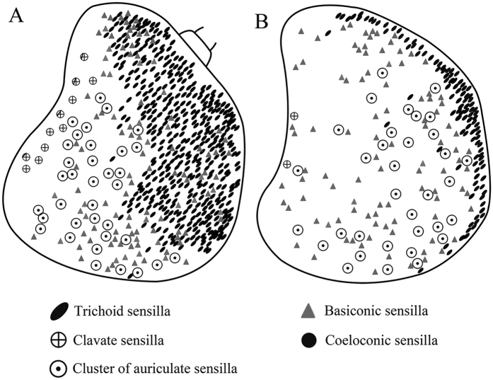
Antennae are among the most elaborate sensory organs in adult flies, and they provide rich information for phylogenic studies. The antennae of five out of eight species of Gasterophilus Leach (G. haemorrhoidalis (Linnaeus), G. intestinalis (De Geer), G. nasalis (Linnaeus), G. nigricornis (Loew) and G. pecorum (Fabricius)), were examined using scanning electron microscopy. The general morphology, including distribution, type, size, and ultrastructure of antennal sensilla were presented, and the definition of auriculate sensilla and sensory pits were updated and clarified. Eighteen antennal characters were selected to construct the first species-level phylogeny of this genus. The monophyly of Gasterophilus was supported by the presence of coeloconic sensilla III on the antennal arista. The species-level cladogram showed G. pecorum branching off at the base, and the remaining species forming the topology (G. intestinalis+ (G. haemorrhoidalis+ (G. nasalis+ G. nigricornis))). Our research shows the importance of the antennal ultrastructure as a reliable source for phylogenetic analysis.


Figures

Figure 1
Figure 1. Features on the head of adult Gasterophilus pecorum.
(A) Antennae located centrally between compound eyes. (B) Anterodorsal surface, dorsolateral margin and posteroventral surface of antenna in resting position. (C) Antennal scape and pedicel. (D) Base and (inset) middle parts of setae on antennal pedicel. (E) Flagellum. (FH) Base, middle and tip of arista. Scale bars: A = 1 mm, B = 500 μm, C = 200 μm, D = 100 μm, 5 μm in inset, E = 250 μm, F = 50 μm, G, H = 10 μm. Abbrevations: Ad, anterodorsal surface; Ar, arista; Br, bristle; Dl, dorsolateral margin; Fn, funiculus; Pd, pedicel; Pv, posteroventral surface; Sc, scape.
Figure 2
Figure 2. Features on the head of adult Gasterophilus nasalis.
(A) Antennae located centrally between compound eyes. (B) Antenna in resting position, showing anterodorsal surface, dorsolateral margin in front and posteroventral surface. (C) Antennal scape. (D) Antennal pedicel. (E) Bristle (mechanoreceptor) on antennal pedicel. (F) Flagellum. Scale bars: A = 1 mm, B, F = 250 μm, C = 200 μm, D = 150 μm, E = 50 μm. Abbrevations: Ad, anterodorsal surface; Ar, arista; Br, bristle; Dl, dorsolateral margin; Fn, funiculus; Pd, pedicel; Pv, posteroventral surface; Sc, scape.
Figure 3
Figure 3. Features on the head of adult Gasterophilus intestinalis.
(A) Antennae located centrally between compound eyes. (B) Antenna in resting position, showing anterodorsal surface, dorsolateral margin in front and posteroventral surface. (C) Antennal scape and pedicel. (D) Bristle (mechanoreceptor) on antennal pedicel. (E) Anterodorsal surface of antennal funiculus and (inset) aristal tip. Scale bars: A, B = 1mm, C = 200 μm, D = 25 μm, E = 250 μm, 50 μm in inset. Abbrevations: Ad, anterodorsal surface; Ar, arista; Br, bristle; Dl, dorsolateral margin; Fn, funiculus; Pd, pedicel; Pv, posteroventral surface; Sc, scape. SP, sensory pit.
Figure 4
Figure 4. Features on the head of adult Gasterophilus haemorrhoidalis.
(A) Antennae located centrally between compound eyes. (B) Antenna in resting position, showing anterodorsal surface, dorsolateral margin in front and posteroventral surface. (C) Antennal scape and pedicel. (D) Bristle (mechanoreceptor) on antennal pedicel. Scale bars: A = 1 mm, B = 0.5 mm, C = 100 μm, D = 50 μm, 20 μm in inset. Abbrevations: Ad, anterodorsal surface; AR, annular ridge; Ar, arista; Br, bristle; C, cone; Dl, dorsolateral margin; Fn, funiculus; PB, pedicellar button; Pd, pedicel; Pv, posteroventral surface; Sc, scape.
Figure 5
Figure 5. Details of antennal pedicel after removal of antennal funiculus and (inset) magnification of pedicellar button.
(A) Gasterophilus pecorum. (B) G. nasalis. (C) G. intestinalis. (D) G. haemorrhoidalis. Scale bars: A = 50 μm, 15 μm in inset, B, C = 50 μm, 10 μm in inset, D = 50 μm, 5 μm in inset. Abbreviations: PB, pedicellar button; AR, annular ridge C, cone.
Figure 6
Figure 6. Scanning electron micrographs of trichoid sensilla and basiconic sensilla on antennal funiculus in Gasterophilus pecorum and G. nasalis.
(A) Trichoid sensilla in G. pecorum. B Basiconic sensilla I in G. pecorum. C Basiconic sensilla II in G. pecorum. D Trichoid sensilla in G. nasalis. E Basiconic sensilla I in G. nasalis. F Basiconic sensilla II in G. nasalis. Scale bars: A, B, E, F = 5 μm, C, D = 10 μm. Abbreviations: Ba, basiconic sensilla; Ba I, basiconic sensilla I; Ba II, basiconic sensilla II; Mt, microtrichia; Tr, trichoid sensilla.
Figure 7
Figure 7. Scanning electron micrographs of trichoid sensilla and basiconic sensilla on antennal funiculus in Gasterophilus intestinalis and G. haemorrhoidalis.
(A) Trichoid sensilla in G. intestinalis. (B) Basiconic sensilla I in G. intestinalis. (C) Basiconic sensilla II in a sensory pit in G. intestinalis. (D) Trichoid sensilla in G. haemorrhoidalis. E Basiconic sensilla I in G. haemorrhoidalis. F Basiconic sensilla II in G. haemorrhoidalis. Scale bars: A, B, C, D, E = 5 μm, F = 2.5 μm. Abbreviations: Ba, basiconic sensilla; Ba I, basiconic sensilla I; Ba II, basiconic sensilla II; Tr, trichoid sensilla.
Figure 8
Figure 8. Scanning electron micrographs of coeloconic sensilla on antennal funiculus and arista in Gasterophilus pecorum and G. nasalis.
(A) Coeloconic sensilla I in G. pecorum. (B) Coeloconic sensilla II in G. pecorum. (C) Coeloconic sensilla III on basal part of arista in G. pecorum. (D) Magnification of coeloconic sensilla III in C. (E) Coeloconic sensilla III in depression on antennal funiculus in G. nasalis. (F) Coeloconic sensilla III on basal part of arista in G. nasalis. (G) Coeloconic sensilla III in F. Scale bars: A, B, E = 2.5 μm, C = 30 μm, D, G = 5 μm, F = 25 μm. Abbreviations: Co I, coeloconic sensilla I; Co II, coeloconic sensilla II; Co III, coeloconic sensilla III.
Figure 9
Figure 9. Scanning electron micrographs of coeloconic sensilla on antennal funiculus and arista in Gasterophilus intestinalis and G. haemorrhoidalis.
(A) Coeloconic sensilla I within a sensory pit in G. intestinalis. (B) Coeloconic sensilla III on antennal arista in G. intestinalis. (C) Coeloconic sensilla III on basal part of arista in G. intestinalis. (D) An coeloconic sensillum I in G. haemorrhoidalis. (E) Coelocronic sensilla III on antennal arista in G. haemorrhoidalis. (F) Coeloconic sensilla III on basal part of arista in G. haemorrhoidalis. Scale bars: A, B, D = 5 μm, C = 20 μm, E, F = 10 μm. Abbreviations: Co I, coeloconic sensilla I; Co III, coeloconic sensilla III.
Figure 10
Figure 10. Scanning electron micrographs of clavate sensilla on antennal funiculus in Gasterophilus nasalis and G. intestinalis.
(A) Clavate sensillum in shallow depression in G. nasalis. (B) Clavate sensilla within a sensory pit in G. nasalis. (C) Clavate sensillum in shallow depression in G. intestinalis. (D) Clavate sensillum in shallow depression in G. haemorrhoidalis. Scale bars: A, C = 2.5 μm, B, D = 5 μm. Abbreviation: Cl, clavate sensilla.
Figure 11
Figure 11. Scanning electron micrographs of auriculate sensilla on antennal funiculus and microtrichiae.
(A) Auriculate sensillum in Gasterophilus nasalis. (B) Auriculate sensillum in G. intestinalis. (C) Auriculate sensilla clustered in a sensory pit in G. haemorrhoidalis. (D) Microtrichiae on antennal funiculus in G. pecorum. (E) Microtrichiae on antennal funiculus in G. nasalis. (F) Microtrichiae on antennal funiculus in G. intestinalis. (G) Microtrichiae on antennal funiculus in G. haemorrhoidalis. Scale bars: A, C = 5 μm, B = 2.5 μm, D = 20 μm, E, F, G = 10 μm, F = 10 μm, G = 10 μm. Abbreviations: Au, auriculate sensilla; Mt, microtrichia.
Figure 12
Figure 12
Distribution of four types of sensilla on anterodorsal surface and dorsolateral margin (A male, C female) and posteroventral surface (B male, D female) of antennal funiculus in Gasterophilus pecorum.
Figure 13
Figure 13
Distribution of four types of sensilla on anterodorsal surface and dorsolateral margin (A male, C female) and posteroventral surface (B male, D female) of antennal funiculus in Gasterophilus nasalis.
Figure 14
Figure 14
Distribution of four types of sensilla on anterodorsal surface and dorsolateral margin (A male, C female) and posteroventral surface (B male, D female) of the antennal funiculus in Gasterophilus intestinalis.
Figure 15
Figure 15
Distribution of four types of sensilla on anterodorsal surface and dorsolateral margin (A male, C female) and posteroventral surface (B male, D female) of antennal funiculus in Gasterophilus haemorrhoidalis.
Figure 16
Figure 16
Distribution of four types of sensilla on anterodorsal surface and dorsolateral margin (A) and posteroventral surface (B) of antennal funiculus in female Gasterophilus nigricornis.
Figure 17
Figure 17. Scanning electron micrographs of sensory pits.
(A) Overview of sensory pits on funiculus in Gasterophilus haemorrhoidalis. (B) Dorsal view of a sensory pit with a fringe of microtrichiae in G. pecorum. (C) Dorsal view of a sensory pit with a fringe of microtrichiae in G. nasalis. (D) Lateral view of a sensory pit with broken microtrichiae. Scale bars: A = 50 μm, B–D = 10 μm.
Figure 18
Figure 18. Consensus tree from 2 most parsimonious trees obtained using implicit enumeration, with characters treated as non-additive and equal weighting.
Figure 19
Figure 19. Cladogram obtained using implicit enumeration, with characters treated as non-additive and under implied weighting (k ≥ 1); or with characters treated as additive and under both equal weighting and implied weighting (k ≥ 1) (length = 33 steps; consistency index CI = 69; retention index RI = 72).
Numbers refer to characters (above branches) and states (below branches) as discussed in the text and in Appendix 1, black circles represent unique character state changes, white circles represent homoplasious character state changes. Underlined numbers are Bremer support values obtained with characters treated as additive using the implicit enumeration analysis.
Figure 20
Figure 20
Three rooted cladograms constructed using maximum likelihood analysis of 28S rDNA sequences (A), COI sequences (B) and 16S rRNA sequences (C) (modified from Otranto et al.58).

Similar articles

Cited by

References

    1. Giroux M., Pape T. & Wheeler T. A. Towards a phylogeny of the flesh flies (Diptera: Sarcophagidae): morphology and phylogenetic implications of the acrophallus in the subfamily Sarcophaginae. Zool J Linn Soc-Lond 158, 740–778 (2010).
    1. Lambkin C. L. et al.. The phylogenetic relationships among infraorders and superfamilies of Diptera based on morphological evidence. Syst Entomol 38, 164–179 (2012).
    1. Yeates D. K. & Wiegmann B. M. Phylogeny and evolution of Diptera: recent insights and new perspectives. In: The evolutionary biology of flies (eds Yeates D. K. & Wiegmann B. M.) 14–44 (Columbia University Press, 2005).
    1. Araujo D. P., Tuan M. J. M., Yew J. Y. & Meier R. Analysing small insect glands with UV-LDI MS: high-resolution spatial analysis reveals the chemical composition and use of the osmeterium secretion in Themira superba (Sepsidae: Diptera). J Evolution Biol 27, 1744–1750 (2014). - PubMed
    1. Wipfler B., Courtney G. W., Craig D. A. & Beutel R. G. First μ-CT-based 3D reconstruction of a dipteran larva—the head morphology of Protanyderus (Tanyderidae) and its phylogenetic implications. J Morphol 273, 968–980 (2012). - PubMed

Publication types

LinkOut - more resources