Review
. 2017 Jan;18(1):5-17.
doi: 10.1038/nrm.2016.121. Epub 2016 Oct 5.

Reverse-topology membrane scission by the ESCRT proteins

Affiliations
Review

Reverse-topology membrane scission by the ESCRT proteins

Johannes Schöneberg et al. Nat Rev Mol Cell Biol. 2017 Jan.

Abstract

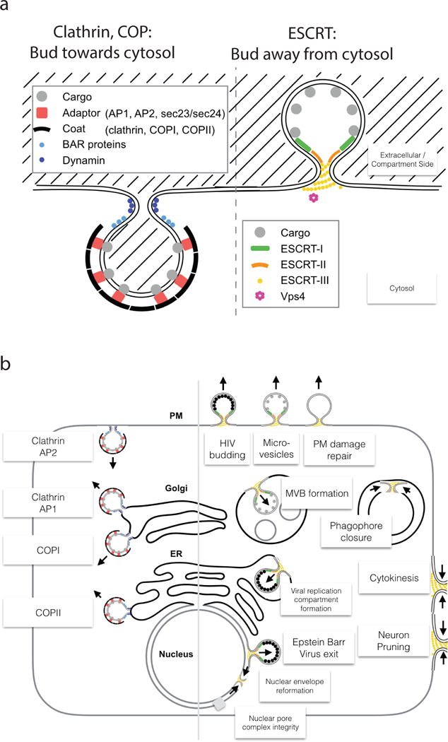
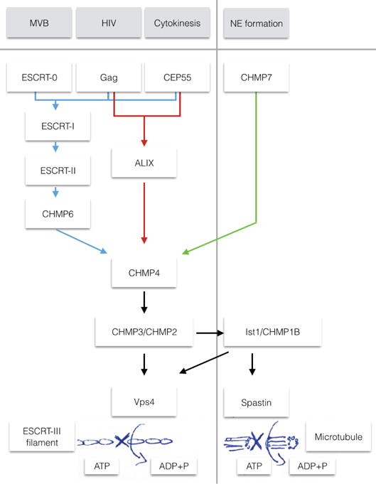
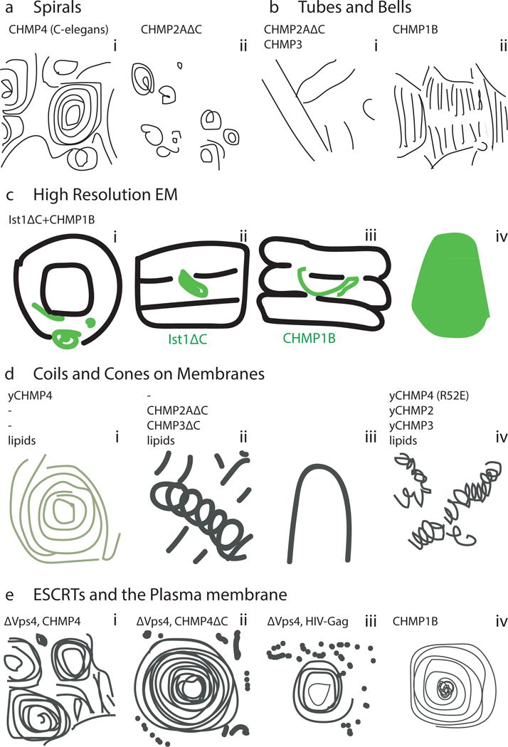
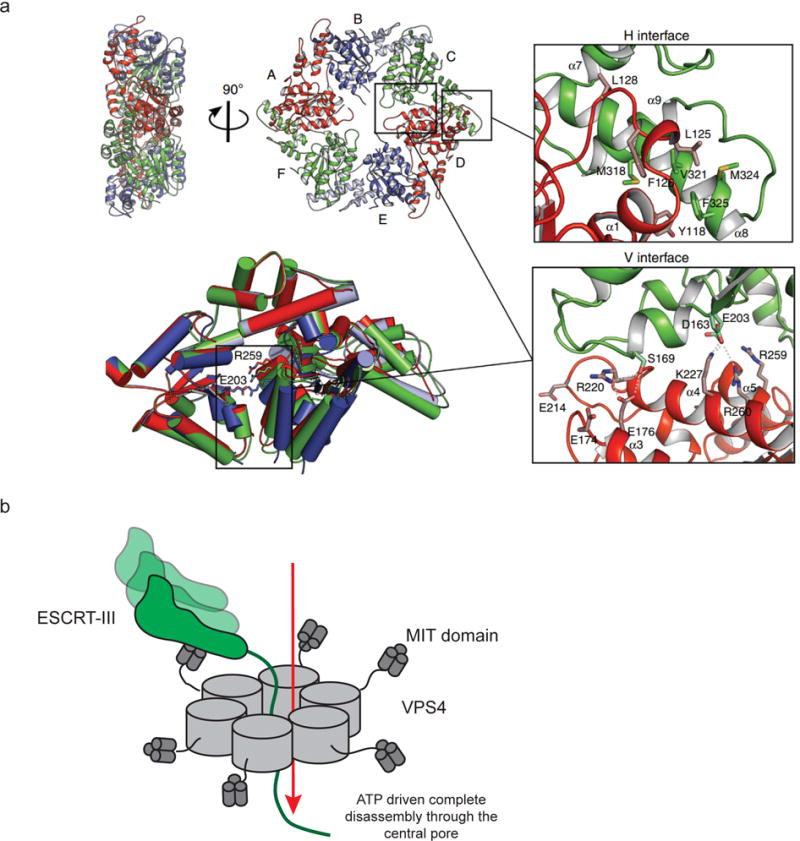
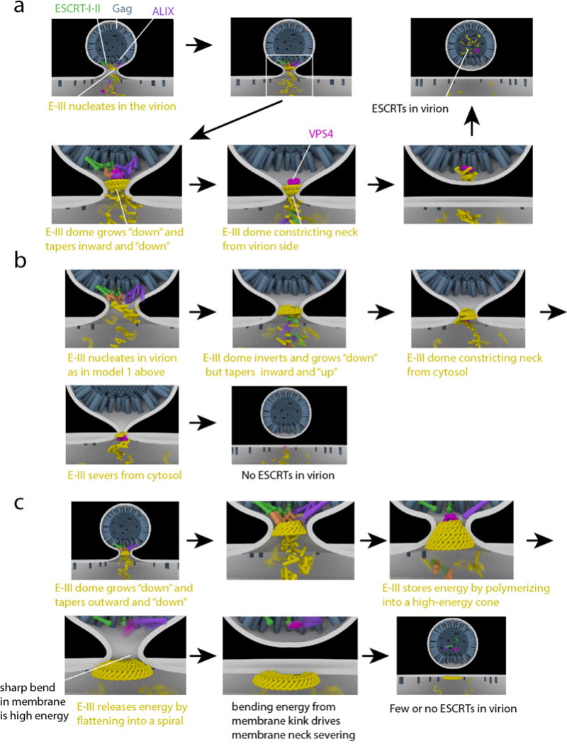
The narrow membrane necks formed during viral, exosomal and intra-endosomal budding from membranes, as well as during cytokinesis and related processes, have interiors that are contiguous with the cytosol. Severing these necks involves action from the opposite face of the membrane as occurs during the well-characterized formation of coated vesicles. This 'reverse' (or 'inverse')-topology membrane scission is carried out by the endosomal sorting complex required for transport (ESCRT) proteins, which form filaments, flat spirals, tubes and conical funnels that are thought to direct membrane remodelling and scission. Their assembly, and their disassembly by the ATPase vacuolar protein sorting-associated 4 (VPS4) have been intensively studied, but the mechanism of scission has been elusive. New insights from cryo-electron microscopy and various types of spectroscopy may finally be close to rectifying this situation.


Figures

Figure 1
Figure 1. Reverse topology membrane scission
a| “Normal” and “reverse” topology membrane scission: “Normal” scission such as occurs in clathrin and coated vesicle biogenesis, whereas “reverse” scission carried out by ESCRTs acts in vesicle budding away from the cytosol. Note that a fundamental difference arises from only the cytosolic membrane side being accessible for protein scaffolding and scission machinery. b| Functions of the ESCRT pathway (right) compared with “normal” scission functions (left). Clathrin, COPI, and COPII are vesicle coats, while AP-1 and AP-2 are adaptor complexes that connect clathrin to membranes and vesicular cargo.
Figure 2
Figure 2. Organization of the ESCRT system
Schematic of the three branches of the ESCRT pathway, explored in the four functions of multivesicular body (MVB) genesis, HIV budding, Cytokinesis and nuclear envelope (NE) formation. The first three of these functions can all use the ‘classic’ pathway (ESCRT-I, ESCRT-II, CHMP6), whereas HIV budding and cytokinesis have respective adaptors that can use ALIX to bridge directly to CHMP4. In NE formation, CHMP7 functions as the bridge to CHMP4. From CHMP4, the canonical pathway uses CHMP2 and CHMP3 that recruit Vps4 for eventual disassembly of the ESCRT filaments. In NE formation, the ESCRT protein IST1 is required, which in turn recruits the microtubule severing enzyme spastin.
Figure 3
Figure 3. The ESCRT-I/II and ALIX branches of the upstream ESCRTs
In nearly every context in which the ESCRTs occur, the upstream components recruit, activate, and organize the polymerization of ESCRT-III. The only exception is in reformation and quality control of the NE, the role of ESCRT-II is apparently replaced by CHMP7. This figure shows how the upstream elements ESCRT-I/II and ALIX organize ESCRT-III at the nanoscale (a) and atomic scales (b and c). a| Struts of putative ESCRT-I/II complexes imaged at a HIV-1 Gag budding site by DEEM (reprinted with permission) (this panel not in NIHMS version due to third party rights). b| ESCRT-II (green) initiates two CHMP4 filaments (blue) via CHMP6 (teal). Structures shown are from RCSB entries 1U5T, 3HTU, and 3JCI. The structures of polymerized IST1 and CHMP1B are used as stand-ins for the CHMP6:CHMP4 complex, whose structure is not known. c| A dimer of ALIX (green) initiates two CHMP4 filaments (blue) via direct interactions with the C-terminus of CHMP4. Structures shown are from RCSB entries 2OEV, 3C3O, and 5FD7.
Figure 4
Figure 4. The ESCRT-III proteins
a| Primary structural organization of the 12 human ESCRT-III proteins. Yeast Snf7 (CHMP4), CHMP2B and CHMP1B are depicted as representatives of the CHMP4A-C, CHMP2A-B, and CHMP1A-B families. The main structural helices are colored: α1 (purple), α2 (blue), α3 (cyan), α4 (green), αA (dark green), αB (dark green), autoinhibitory helix α5 (red) and interaction helix α6 (yellow). The C-termini of ESCRT-IIIs carry microtubule-interacting and transport (MIT)-interacting motifs (MIM1, MIM2) that are responsible for binding to other components of the ESCRT pathway, such as Vps4. b| Crystal structures of ESCRT-III monomers. Color coding same as a. CHMP1B and CHMP4 have both been found to adopt open conformations, with helices α2 and α3 merging, . Ist1ΔC and CHMP3 are shown in the closed conformations in which they have been crystallized, .
Figure 5
Figure 5. ESCRT-III assembly structures
a| Spirals. i) Cryo-EM of C. elegans CHMP4. ii) Negative-stain EM of CHMP2AΔC polymers. Scale bar: 40nm. b| Tubes and bell shapes. i) Cryo-EM of CHMP2AΔC–CHMP3 tubes with cones (*) and tubes (∧). Scale bar: 40nm. ii) Cryo-EM of CHMP1B. c| Cryo-EM structure of ESCRT-III tubular assembly. i) End-on view of IST1ΔC-CHMP1B tube with IST1ΔC in light green and CHMP1B in dark green. ii) External view of the reconstructed helix with IST1ΔC highlighted. iii) Cutaway view with CHMP1B highlighted. iv) Side view of an IST1ΔC-CHMP1B cone. Scale bar: 5.1nm. d| EM of ESCRT-III assemblies on membranes. i) TEM of a single Snf7 spiral on a POPC:POPS membrane. ii) CHMP2AΔC-CHMP3ΔC tubes in the presence of SOPC:DOPS. Tube diameter ~55nm. iv) Snf7R52E:Vps24:Vps2 2:1:1 helices assembled on POPC:POPC:PI(3)P monolayers. e| EM of ESCRT-III in cells. i) Anaglyph of plasma membranes from COS-7 cells expressing CHMP4A. Scale bar: 100nm. ii) Filament spirals on COS-7 cell membranes expressing CHMP4A1-164. Scale bar: 100nm. iii) HEK293T cells treated with Vps4A & Vps4B siRNA accumulate filament-encircled Gag assemblies. Scale bar: 100nm. iv) Spirals on COS-7 cells expressing FLAG-CHMP1B. Scale bar: 50nm. (this figure not included in NIHMS version due to third party rights)
Figure 6
Figure 6. ESCRT complex disassembly
a| The structure of MsVps4 shows how pore structure, nucleotide binding, and hexamerization are interconnected. Crystal structure of the MsVps4ΔMIT pseudohexamer viewed from two orientations. The protomers are labeled from A to F and identical protomers are represented in the same color, except for protomers B and E, whose small ATPase domain is shown in light blue and the large ATPase domain in dark blue. Superposition of the three different dimers present in the pseudohexamer and molecular interactions at the two different interfaces are shown. b| VPS4 hexamers completely disassemble target ESCRT-III subunits by disassembling them through the central pore.
Figure 7
Figure 7. Models for ESCRT-mediated scission
HIV-1 Gag (blue) accumulates at the membrane (white), causing initial membrane deformation. ESCRT-I/ESCRT-II (green/orange) and ALIX (purple) are recruited by Gag. ESCRT-III (yellow) is recruited by ESCRT-II and ALIX, and polymerizes in the bud neck. a| “Original” dome model. ESCRT-III polymerizes away from the virion and towards the cytosol whilst forming consecutively narrower rings such that the dome tapers to a point in the same direction in which it grows. VPS4 (pink) depolymerizes ESCRT-III and scission occurs. ESCRTs are retained in the virion. b| Reverse dome model. ESCRTs are released to the cytosol. As compared to the “original” dome model (a), ESCRT-III grows in the same direction, but tapers in the opposite direction. It is not clear how this paradoxical mode of growth could occur in practice, but it would presumably require active remodeling by VPS4. c| Buckling model. As in the reverse dome model (b), ESCRTs are released to the cytosol at the end. ESCRT-III polymerizes outward from the virion towards the cytosol, with consecutive wider rings. The cone is higher in energy than a flat spiral. Conversion of the cone to a spiral releases the tension, but at the cost of creating sharp bends where the virion is attached to the plasma membrane. The high energy of these bends is released when the virion is severed.

References

    1. Votteler J, Sundquist WI. Virus Budding and the ESCRT Pathway. Cell Host Microbe. 2013;14:232–241. - PMC - PubMed
    1. Martin-Serrano J, Neil SJD. Host factors involved in retroviral budding and release. Nature Reviews Microbiology. 2011;9:519–531. - PubMed
    1. Olmos Y, Carlton JG. The ESCRT machinery: new roles at new holes. Curr Opin Cell Biol. 2016;38:1–11. - PMC - PubMed
    1. Campsteijn C, Vietri M, Stenmark H. Novel ESCRT functions in cell biology: spiraling out of control? Curr Opin Cell Biol. 2016;41:1–8. - PubMed
    1. Hurley JH. ESCRTs are everywhere. EMBO J. 2015;34:2398–2407. - PMC - PubMed

MeSH terms

Substances