Review
. 2016 Sep 20:7:168.
doi: 10.3389/fgene.2016.00168. eCollection 2016.

Gender Differences in Adipocyte Metabolism and Liver Cancer Progression

Affiliations
Review

Gender Differences in Adipocyte Metabolism and Liver Cancer Progression

Otto K-W Cheung et al. Front Genet. .

Abstract

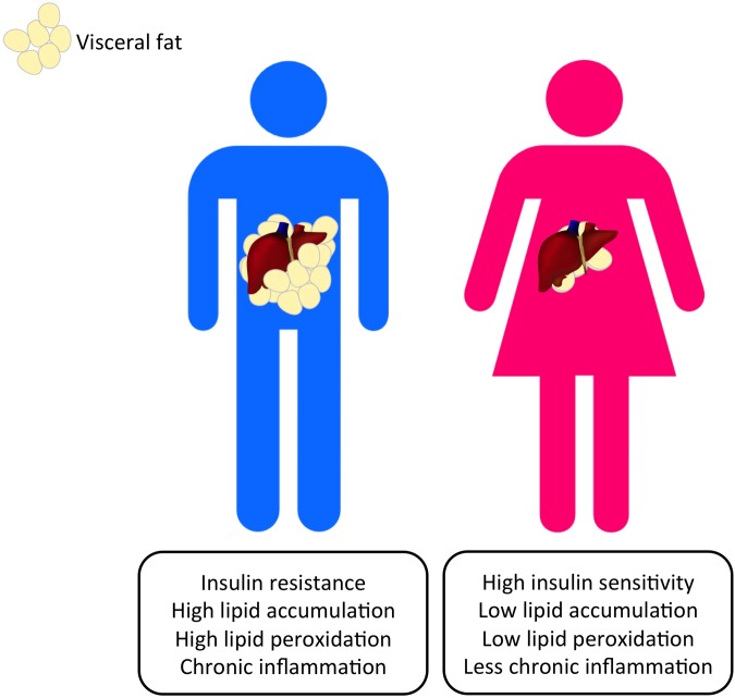
Liver cancer is the third most common cancer type and the second leading cause of deaths in men. Large population studies have demonstrated remarkable gender disparities in the incidence and the cumulative risk of liver cancer. A number of emerging risk factors regarding metabolic alterations associated with obesity, diabetes and dyslipidemia have been ascribed to the progression of non-alcoholic fatty liver diseases (NAFLD) and ultimately liver cancer. The deregulation of fat metabolism derived from excessive insulin, glucose, and lipid promotes cancer-causing inflammatory signaling and oxidative stress, which eventually triggers the uncontrolled hepatocellular proliferation. This review presents the current standing on the gender differences in body fat compositions and their mechanistic linkage with the development of NAFLD-related liver cancer, with an emphasis on genetic, epigenetic and microRNA control. The potential roles of sex hormones in instructing adipocyte metabolic programs may help unravel the mechanisms underlying gender dimorphism in liver cancer and identify the metabolic targets for disease management.

Keywords: adipocyte; adipokine; adiponectin; epigenetic; gender dimorphism; hepatocellular carcinoma; leptin; metabolism.


Figures

FIGURE 1
FIGURE 1
Gender dimorphism occurs in the altered metabolic pathways that can lead to the progression of HCC. Visceral adiposity can be observed more commonly in men and nurtures a pro-carcinogenic microenvironment that promotes HCC initiation.
FIGURE 2
FIGURE 2
Visceral adiposity increases the release of pro-inflammatory adipokines and reduces the protective counterpart, causing immune cell infiltration, liver injury, and the subsequent pathological syndromes. The increase in the release of FFAs and glycerol to the liver from visceral fat also promotes lipid peroxidation and thus increases HCC risks. MDSCs: myeloid-derived suppressor cells.
FIGURE 3
FIGURE 3
Protective single nucleotide polymorphisms (SNPs) in ADIPOQ and LEP genes that lead to differential serum adiponectin and leptin levels occur frequently in females. A respective higher adiponectin and a lower leptin level can be found in female subjects. SNPs in ADIPOQ and LEP genes potentially reduce HCC risks and development in women through inflammatory and oncogenic pathways.
FIGURE 4
FIGURE 4
Hypermethylation of LEP promoter in female offspring offers a protective effect against maternal high-fat diets by lowering leptin production in mice. Maternal high-fat diets affect offspring’s epigenetic regulation on the LEP gene. Obesogenic environments during the pre-conceptional period and the early phase of development led to a lower methylation state of the LEP promoter in male rodent offspring than their female counterparts. The higher leptin production in male offspring was also associated with overweight, insulin resistance and hepatic steatosis, which could help explain the gender dimorphism in leptin production and the development of liver diseases and HCC.

References

    1. Allard C., Desgagné V., Patenaude J., Lacroix M., Guilemette L., Battista M. C., et al. (2015). Mendelian randomization supports causality between maternal hyperglycemia and epigenetic regulation of leptin gene in newborns. Epigenetics 10 342–351. 10.1080/15592294.2015.1029700 - DOI - PMC - PubMed
    1. Ambati S., Yu P., Mckinney E. C., Kandasamy M. K., Hartzell D., Baile C. A., et al. (2016). Adipocyte nuclei captured from VAT and SAT. BMC Obes. 3:35 10.1186/s40608-016-0112-6 - DOI - PMC - PubMed
    1. Arner P., Sinha I., Thorell A., Rydén M., Dahlman-Wright K., Dahlman I. (2015). The epigenetic signature of subcutaneous fat cells is linked to altered expression of genes implicated in lipid metabolism in obese women. Clin. Epigen. 7:93 10.1186/s13148-015-0126-9 - DOI - PMC - PubMed
    1. Baffy G., Brunt E. M., Caldwell S. H. (2012). Hepatocellular carcinoma in non-alcoholic fatty liver disease: an emerging menace. J. Hepaotol. 56 1384–1391. 10.1016/j.jhep.2011.10.027 - DOI - PubMed
    1. Barros R. P., Gabbi C., Morani A., Warner M., Gustafsson J. A. (2009). Participation of ERα and ERβ in glucose homeostasis in skeletal muscle and white adipose tissue. Am. J. Physiol. Endocrinol. Metab. 297 E124–E133. 10.1152/ajpendo.00189.2009 - DOI - PubMed

LinkOut - more resources