

Danger zone analysis using cone beam computed tomography after apical enlargement with K3 and K3XF in a manikin model
- PMID: 27703602
- PMCID: PMC5045681
- DOI: 10.4317/jced.52523
Danger zone analysis using cone beam computed tomography after apical enlargement with K3 and K3XF in a manikin model
Abstract
Background: The objective of the study was to evaluate and compare how apical enlargement with K3 and K3XF nickel-titanium (NiTi) rotary instruments reduces the root thickness in the danger zone and affects canal transportation and centering ability in mandibular molar mesial canals in a manikin extracted tooth model.
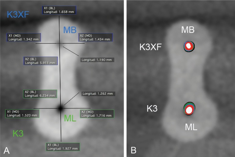
Material and methods: Seventy-two mesial root canals of first mandibular molars were instrumented. Initial and post-instrumentation Cone Beam Computed Tomography scans were performed after root canal preparation up to size 25, 30, 35 and 40 files. Canal transportation, canal centering and remaining root dentin thickness toward the danger zone were calculated in sections 1, 2 and 3 mm under the furcation level. Data were analyzed using non-parametric Kruskal-Wallis analysis of variance at a significance level of P < 0.05.
Results: K3 instruments removed more dentin toward the danger zone compared with K3XF instruments (P< .05) and significant differences in dentin thickness were found when canal enlargement was performed to a #35-40 with both systems (P< 0.05). No significant differences in canal transportation and centering ability were found between systems, except when canal enlargement was performed to a #40 (P = 0,0136). No differences were observed when comparing the number of uses in both systems (P> 0.05).
Conclusions: Under the conditions of this study K3 removed a significant amount of dentin at the furcation level compared with the R-Phase K3XF rotary system in curved root canals. Enlargement to a 35-40/04 file removed significantly more dentin with both systems. Key words:K3, K3XF, R-phase, center ability, canal transportation, dentin thickness, increased apical enlargement, danger zone, dentin thickness.
Conflict of interest statement
The authors deny any conflicts of interest.
Figures
References
-
- Abou-Rass M, Frank AL, Glick DH. The anticurvature filing method to prepare the curved root canal. J Am Dent Assoc. 1980;101:792–4. - PubMed
-
- Bower RC. Furcation morphology relative to periodontal treatment (furcation root surface anatomy) J Periodontol. 1979;50:366–74. - PubMed
-
- Kessler JR, Peters DD, Lorton L. Comparison of the relative risk of molar root perforation using various endodontic instrumentation techniques. J Endod. 1983;9:439–77. - PubMed
-
- Garcia Filho PF, Letra P, Menezes R, Rezende do Carmo AM. Danger zone in mandibular molars before instrumentation: An in vitro study. J Appl Oral Sci. 2003;11:324–6. - PubMed
-
- Berutti L, Fedon G. Thickness of cementum/dentin in mesial root of mandibular first molars. J Endod. 1992;18:545–8. - PubMed
LinkOut - more resources
Full Text Sources
Other Literature Sources