. 2017 Jan;125(1):38-45.
doi: 10.1111/apm.12597. Epub 2016 Oct 5.

Early implant-associated osteomyelitis results in a peri-implanted bacterial reservoir

Affiliations

Early implant-associated osteomyelitis results in a peri-implanted bacterial reservoir

Louise Kruse Jensen et al. APMIS. 2017 Jan.

Abstract

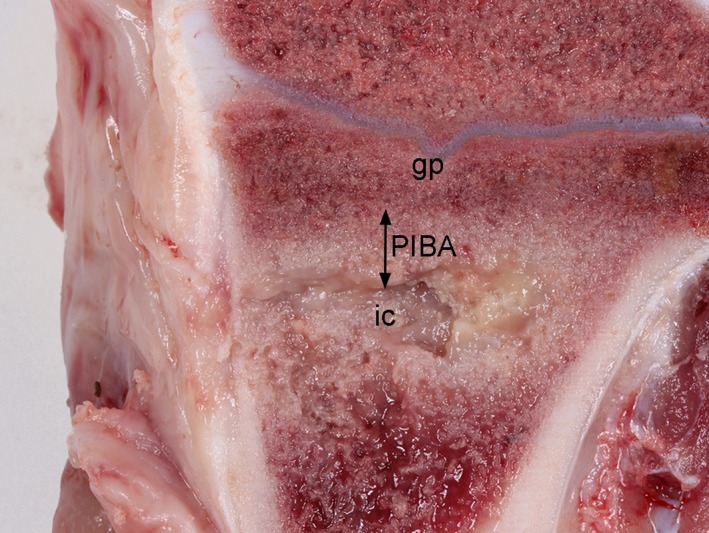
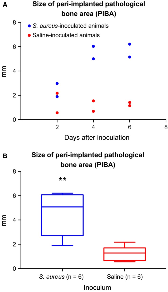
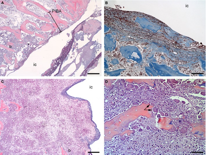
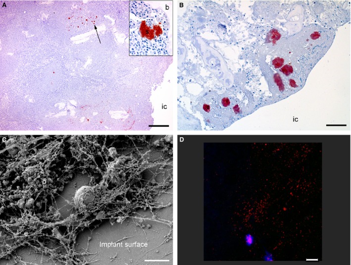
Implant-associated osteomyelitis (IAO) is a common complication in orthopedic surgery. The aim of this study was to elucidate how deep IAO can go into the peri-implanted bone tissue within a week. The study was performed in a porcine model of IAO. A small steel implant and either 104 CFU/kg body weight of Staphylococcus aureus or saline was inserted into the right tibial bone of 12 pigs. The animals were consecutively killed on day 2, 4 and 6 following implantation. Bone tissue around the implant was histologically evaluated. Identification of S. aureus was performed immunohistochemically on tissue section and with scanning electron microscopy and peptide nucleic acid in situ hybridization on implants. The distance of the peri-implanted pathological bone area (PIBA), measured perpendicular to the implant, was significantly larger in infected animals compared to controls (p = 0.0014). The largest differences were seen after 4 and 6 days of inoculation, where PIBA measurements of up to 6 mm were observed. Positive S. aureus bacteria were identified on implants and from 25 μm to 6 mm into PIBA. This is important knowledge for optimizing outcomes of surgical debridement in osteomyelitis.

Keywords: S. aureus; Implant-associated osteomyelitis; animal experiment; histopathology.


Figures

Figure 1
Figure 1
Macroscopic pathological changes in a porcine tibial bone 6 days after S. aureus inoculation and insertion of a steel implant. The implant was inserted just below the grow plate (gp) in the trabecular bone tissue. The implant has been removed on the picture, presenting the implant cavity (ic). Purulent exudation is seen in the implant cavity and within the surrounding bone tissue. The peri‐implanted bone tissue with pathological changes is referred to as the peri‐implanted pathological bone area (PIBA).
Figure 2
Figure 2
(A) The size in mm of the peri‐implanted pathological bone area (PIBA) in a porcine model of implant‐associated osteomyelitis 2, 4 and 6 days after tibial insertion of a steel implant and injection of Staphylococcus aureus (blue) or saline (red). PIBA was measured perpendicular from the implant cavity and until normal tissue architecture occurred. (B) Grouping of animals inoculated with Staphylococcus aureus and animals inoculated with saline. The figure shows the size in mm of the peri‐implanted pathological bone area (PIBA). Boxes, 95% confidence interval; whiskers, min. and max. value: bold line, mean value. The size of PIBA was significantly (** = P ≤ 0.001) increased in pigs inoculated with S. aureus.
Figure 3
Figure 3
Overview of histopathological bone changes in porcine tibial bones following 2 (A) and 6 (B, C and D) days after S. aureus or saline inoculation and insertion of a steel implant. Pictures A and B represent pigs inoculated with saline, and pictures C and D represent pigs inoculated with S. aureus. IC: implant cavity. Picture A: an overview of the peri‐implanted pathological bone area (PIBA) after 2 days of inoculation. HE, bar = 400. Picture B: a thin area of primarily fibroblasts and collagen was seen on the surface toward the implant cavity. Masson's trichrome, bar = 250. Picture C: a large area of fibroblasts, collagen, giant cells and macrophages was seen toward the implant cavity. Almost no bone (b) tissue was left within PIBA. HE, bar = 300. Picture D: a dominant finding between infected and control pigs after day 4 of inoculation was a substantial presence of active osteoclasts (arrow). HE, bar = 200.
Figure 4
Figure 4
Visualization of bacteria inside bone tissue and on the surface of steel implants from a porcine model of implant‐associated osteomyelitis inoculated with S. aureus or saline. IC: implant cavity. Picture A: six days after bacterial inoculation, S. aureus‐positive bacteria (arrow) were seen in the peri‐implanted pathological bone area (PIBA). Insert; Close‐up of a positive S. aurous colony, IHC staining for S. aureus, bar = 300. Picture B: four days after inoculation with saline, S. aureus‐positive bacteria were seen enclosed just inside PIBA. These bacteria are supposed to be a result of self‐contamination, as the Spa type of the implant cavity revealed another S. aureus strain as the one used for inoculation, IHC staining for S. aureus, bar = 200. Picture C: scanning electron microscopy (SEM) of an implant surface following 4 days of inoculation with S. aureus showing bacteria (b) and extracellular material (e), bar = 5 μm. Picture D: peptide nucleic acid fluorescence in situ hybridization (PNA FISH) of implant surface following 6 days of inoculation with S. aureus. Bacteria positive for the S. aureus probe light up in red and leukocytes in blue, bar = 20.

References

    1. Lew DP, Waldvogel FA. Osteomyelitis. N Engl J Med 1997;336:999–1007. - PubMed
    1. Sendi P, Zimmerli W. Antimicrobial treatment concepts for orthopedic device‐related infections. Clin Microbiol Infect 2012;18:1176–84. - PubMed
    1. Jorgensen PN, Meyer R, Dagnaes‐Hansen F, Fuursted K, Petersen E. A modified chronic infection model for testing treatment of Staphylococcus aureus biofilms on implants. PLoS One 2014;9:1–7. - PMC - PubMed
    1. Krenek L, Farng E, Zingmond D, SooHoo NF. Complication and revision rates following total elbow arthroplasty. J Hand Surg Am 2011;36:68–73. - PubMed
    1. Zimmerli W, Moser C. Pathogenesis and treatment concepts of orthopedic biofilm infections. FEMS Immunol Med Microbiol 2012;65:158–68. - PubMed

MeSH terms

LinkOut - more resources