. 2017 Mar;32(3):522-535.
doi: 10.1002/jbmr.3011. Epub 2016 Oct 27.

Control of Bone Anabolism in Response to Mechanical Loading and PTH by Distinct Mechanisms Downstream of the PTH Receptor

Affiliations

Control of Bone Anabolism in Response to Mechanical Loading and PTH by Distinct Mechanisms Downstream of the PTH Receptor

Jesus Delgado-Calle et al. J Bone Miner Res. 2017 Mar.

Abstract

Osteocytes integrate the responses of bone to mechanical and hormonal stimuli by poorly understood mechanisms. We report here that mice with conditional deletion of the parathyroid hormone (PTH) receptor 1 (Pth1r) in dentin matrix protein 1 (DMP1)-8kb-expressing cells (cKO) exhibit a modest decrease in bone resorption leading to a mild increase in cancellous bone without changes in cortical bone. However, bone resorption in response to endogenous chronic elevation of PTH in growing or adult cKO mice induced by a low calcium diet remained intact, because the increased bone remodeling and bone loss was indistinguishable from that exhibited by control littermates. In contrast, the bone gain and increased bone formation in cancellous and cortical bone induced by daily injections of PTH and the periosteal bone apposition induced by axial ulna loading were markedly reduced in cKO mice compared to controls. Remarkably, however, wild-type (WT) control littermates and transgenic mice overexpressing SOST injected daily with PTH exhibit similar activation of Wnt/β-catenin signaling, increased bone formation, and cancellous and cortical bone gain. Taken together, these findings demonstrate that Pth1r in DMP1-8kb-expressing cells is required to maintain basal levels of bone resorption but is dispensable for the catabolic action of chronic PTH elevation; and it is essential for the anabolic actions of daily PTH injections and mechanical loading. However, downregulation of Sost/sclerostin, previously shown to be required for bone anabolism induced by mechanical loading, is not required for PTH-induced bone gain, showing that other mechanisms downstream of the Pth1r in DMP1-8kb-expressing cells are responsible for the hormonal effect. © 2016 American Society for Bone and Mineral Research.

Keywords: GENETIC ANIMAL MODELS; MOLECULAR PATHWAYS-REMODELING; OSTEOCYTES; PTH/VITD/FGF23; WNT/Β-CATENIN/LRPS.


Conflict of interest statement

Disclosures

All authors state that they have no conflicts of interest.

Figures
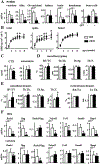
Fig. 1.
Fig. 1.
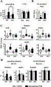
Absence of Pth1r signaling in DMP1-8kb–expressing cells increases cancellous bone mass. (A) Pth1r mRNA expression in whole bone, Ot-enriched bone, large organs, and GFP− and GFP+ primary cells isolated from fl/fl (n=5) and cKO mice (n=5). (B) Longitudinal BMD measurements (2 to 8 months of age) in fl/fl (n=16) and cKO (n=17) female mice. (C) Serum levels of osteocalcin and CTX (n=11 fl/fl and n=17 cKO mice), (D, E) μCT analyses of the spinal/femoral cancellous and femoral mid-diaphysis cortical bone, and (F) gene expression in 4-month-old female mice (n=7 fl/fl and n=7 cKO mice). Bars represent means±SD. *p≤0.05 versus control fl/fl mice, by t test or Mann-Whitney rank sum test (C). Ot=osteocyte; BV/TV=bone volume over tissue volume; Tb.Th=trabecular thickness; Tb.Sp=trabecular separation; Tb.N=trabecular number; BAT/TA=bone area over tissue area; Ct.Th=cortical thickness.
Fig. 2.
Fig. 2.
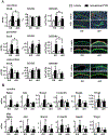
Mice lacking Pth1r in DMP1-8kb–expressing cells exhibit a normal catabolic response to chronic increase of endogenous PTH secretion. (A, B) Serum PTH and CTX levels in growing (1-month-old; normal/deficient Ca diet; n=7/7 fl/fl and n=8/7 cKO female and male mice) and adult (4-month-old; normal/deficient Ca diet; n=9/9 fl/fl and n=7/6 cKO female and male mice) fl/fl and cKO mice fed a normal or calcium-deficient diet. Boxplots of total, spinal, and femoral BMD percent change calculated over initial BMD measurements in growing (C) and adult (D) male and female mice. Middle line in box represents the median, whiskers the 95% confidence interval of the mean, and open circles are outliers from the 95% confidence interval. μCT analysis showing similar cancellous and cortical bone loss in fl/fl and cKO growing (E) and adult mice (F) fed a calcium-deficient diet. Bars represent means±SD. *p≤0.05 versus control fl/fl mice of each group, unless otherwise indicated by the lines, by two-way ANOVA.
Fig. 3.
Fig. 3.
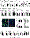
Mice lacking Pth1r in DMP1-8kb–expressing cells display reduced anabolic response to intermittent administration of PTH. (A) Serum osteocalcin and CTX levels in 4-month-old fl/fl and cKO mice after 4 weeks of daily injections of PTH. (B) Sost mRNA expression after treatment with PTH for 24 hours in Ot-enriched bone organ cultures collected from fl/fl and cKO mice (n=3/group fl/fl and n=4/group cKO mice). (C, D) Boxplots of total, spinal, and femoral BMD percent change calculated over initial BMD measurements, and μCT analysis of the cancellous bone in the distal femur and cortical bone in the femoral mid-diaphysis in 4-month-old fl/fl and cKO mice after 4 weeks of daily injections of PTH. Middle line in box represents the median, whiskers the 95% confidence interval of the mean, and open circles are outliers from the 95% confidence interval. Bars represent means±SD. veh/PTH: n=10/8 fl/fl and n=9/8 cKO male and female mice. *p≤0.05 versus control mice receiving vehicle control injections of each group, unless otherwise indicated by the lines, by two-way ANOVA. Ot=osteocyte.
Fig. 4.
Fig. 4.
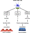
Osteocytic Pth1r signaling is required for PTH-induced stimulation of bone formation and activation of Wnt/β-catenin signaling. (A) Bone dynamic histomorphometric analyses performed on femurs from 4-month-old fl/fl and cKO female mice after 4 weeks of PTH administration. Representative images of the labelling in the cancellous, periosteal, and endocortical bone surfaces are shown. (B) Osteoblast markers (Bglap, Alpl, and Runx2) and Wnt target genes (Cyclin D1, Smad6, and Wisp2) mRNAexpression in cancellous (vertebra) and cortical (tibia diaphysis) bones. Bars represent means±SD. veh/PTH: n=10/8 fl/fl and n=9/8 cKO mice. *p≤0.05 versus control mice receiving vehicle control injections of each group, unless otherwise indicated by the lines, by two-way ANOVA.
Fig. 5.
Fig. 5.
PTH-induced bone gain does not require Sost/Sclerostin downregulation. (A) mRNA expression of endogenous Sost and human transgenic SOST in cortical bone of the tibia diaphysis, (B) serum osteocalcin and CTX levels, (C) boxplots of total, spinal, and femoral BMD percent change calculated over initial BMD measurements (middle line in box represents the median, whiskers the 95% confidence interval of the mean, and open circles are outliers from the 95% confidence interval), (D) and μCT analysis of the cancellous bone in the distal femur and cortical bone in the femoral mid-diaphysis in 4-month-old WT and TG female mice after 4 weeks of PTH administration. Histomorphometric analysis of bone formation dynamic parameters (E), representative images of the labelling in the cancellous bone surfaces (F), and osteoclast numbers/surface and Rankl and Opg mRNA expression (G) in cancellous bone (spine) from WT and TG mice. (H) Osteoblast markers (Bglap, Alpl, and Runx2) and Wnt target genes (Cyclin D1, Smad6, Wisp2, and Cx43) mRNA expression. Bars represent means±SD. veh/PTH: n=10/8 WT and n=11/10 TG mice. *p≤0.05 versus control mice receiving vehicle control injections of each group, unless otherwise indicated by the lines, by two-way ANOVA. §p≤0.05 versus vehicle-treated mice within each genotype by t test. n.d.=not detected; N.Oc/BS=number of osteoclast per bone surface; Oc.S/BS=osteoclast surface per bone surface.
Fig. 6.
Fig. 6.
Deletion of Pth1r in DMP1-8kb–expressing cells markedly impairs the osteogenic response to mechanical loading. (A) Ex vivo strain measurements and linear relation between applied peak force and mechanical strain at the medial surface of the ulnar mid-diaphysis of 4-month-old fl/fl or cKO female mice. n=5 fl/fl and n=6 cKO mice. (B) Bone histomorphometric measurements were performed in the periosteal surface of control and loaded mid-diaphysis ulna from WT and cKO female mice. n=8/group fl/fl and n=5/group cKO mice. (C) Relative bone formation rate, BFR/BS of loaded minus non-loaded ulna for each mouse at each strain magnitude. (D) Sost, Naked, and Cx43 mRNA expression in ulna from fl/fl and cKO mice loaded at high magnitude strain. *p≤0.05 versus fl/fl mice (A and C) or versus non-loaded ulna of the corresponding genotype (B and D), by t test. Means±SD are shown. MS/BS=percent mineralizing bone surface per bone surface; MAR=mineral apposition rate; BFR/BS=bone formation rate per bone surface.
Fig. 7.
Fig. 7.
PTH receptor in DMP1-8kb–expressing cells and bone remodeling. PTH receptor in DMP1-8kb–expressing cells maintains basal levels of resorption under physiological levels of endogenous PTH, whereas it is dispensable for the bone loss induced by continuous elevation of the hormone. Moreover, PTH receptor signaling in DMP1-8kb–expressing cells is required to stimulate Wnt/β-catenin signaling and for a full anabolic response to mechanical loading and intermittent administration of PTH. However, although mechanical loading requires downregulation of Sost/Sclerostin to activate Wnt/β-catenin signaling, other products regulated by the PTH receptor signaling in DMP1-8kb–expressing cells, but different from Sclerostin, are responsible for the stimulation of the pathway.

References

    1. Silva BC, Costa AG, Cusano NE, Kousteni S, Bilezikian JP. Catabolic and anabolic actions of parathyroid hormone on the skeleton. J Endocrinol Invest. 2011;34:801–10. - PMC - PubMed
    1. Saliba W, El-Haddad B. Secondary hyperparathyroidism: pathophysiology and treatment. J Am Board Fam Med. 2009;22:574–81. - PubMed
    1. Ma YL, Cain RL, Halladay DL, et al. Catabolic effects of continuous human pth (1–38) in vivo is associated with sustained stimulation of rankl and inhibition of osteoprotegerin and gene-associated bone formation. Endocrinology. 2001;142:4047–54. - PubMed
    1. Jilka RL. Molecular and cellular mechanisms of the anabolic effect of intermittent PTH. Bone. 2007;40:1434–46. - PMC - PubMed
    1. Ben-Awadh A, Delgado-Calle J, Tu X et al. Parathyroid hormone receptor signaling induces bone resorption in the adult skeleton by directly regulating the RANKL gene in osteocytes. Endocrinology. 2014;155:2797–809. - PMC - PubMed

MeSH terms

Substances