. 2016 Nov 15;7(46):75407-75424.
doi: 10.18632/oncotarget.12249.

Indoleamine 2,3-dioxygenase regulates anti-tumor immunity in lung cancer by metabolic reprogramming of immune cells in the tumor microenvironment

Affiliations

Indoleamine 2,3-dioxygenase regulates anti-tumor immunity in lung cancer by metabolic reprogramming of immune cells in the tumor microenvironment

Cara C Schafer et al. Oncotarget. .

Abstract

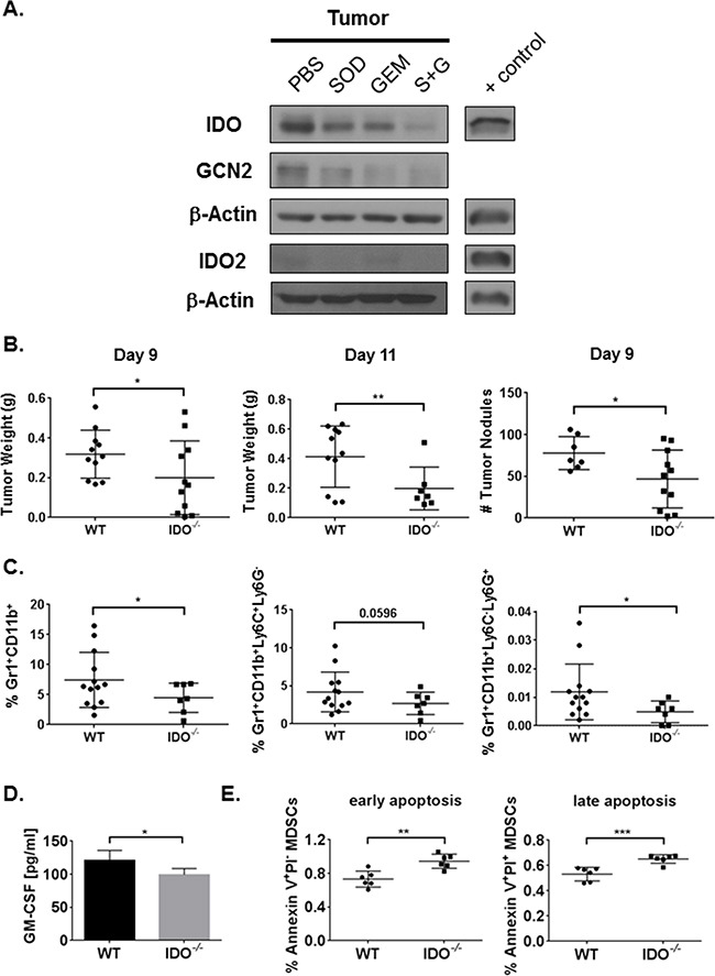
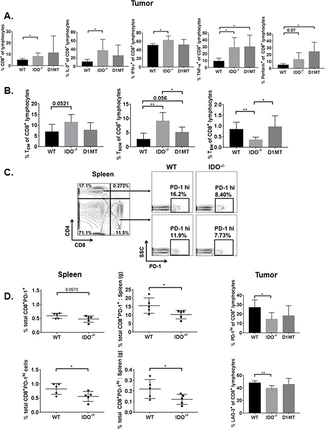
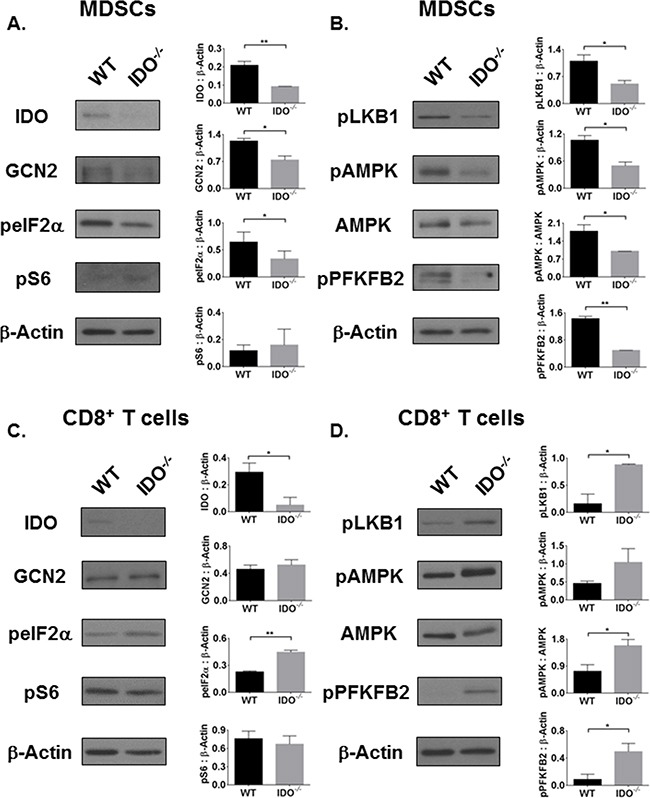
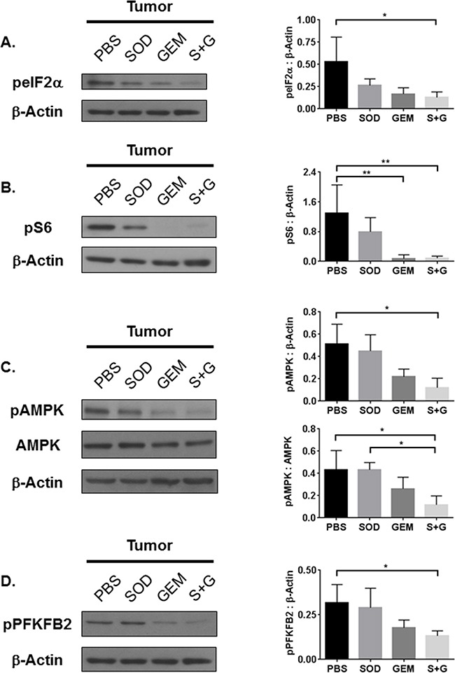
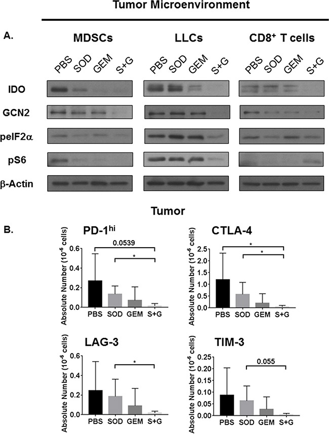
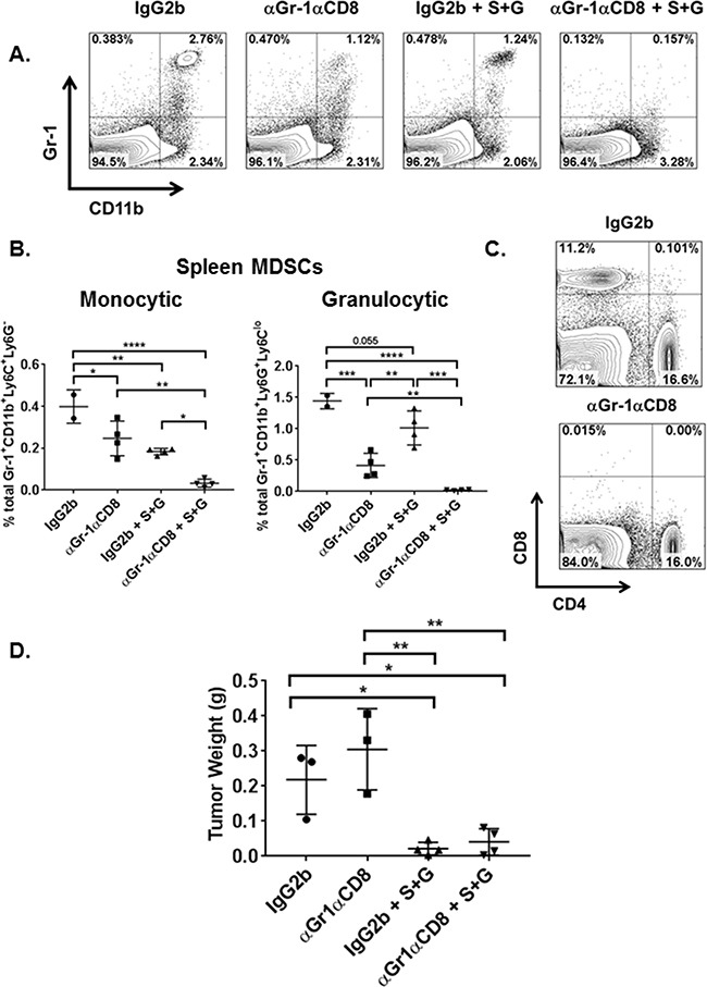
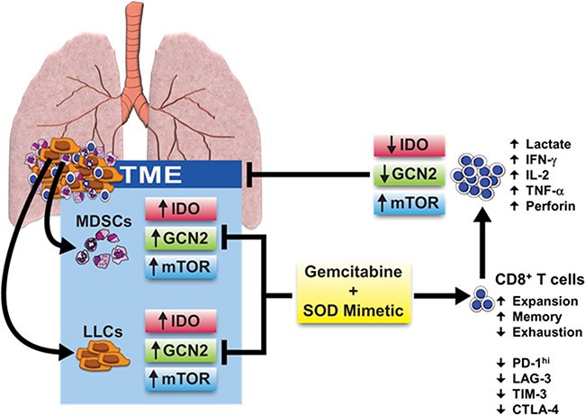
Indoleamine 2,3-dioxygenase (IDO) has been implicated in immune evasion by tumors. Upregulation of this tryptophan (Trp)-catabolizing enzyme, in tumor cells and myeloid-derived suppressor cells (MDSCs) within the tumor microenvironment (TME), leads to Trp depletion that impairs cytotoxic T cell responses and survival; however, exact mechanisms remain incompletely understood. We previously reported that a combination therapy of gemcitabine and a superoxide dismutase mimetic promotes anti-tumor immunity in a mouse model of lung cancer by inhibiting MDSCs, enhancing polyfunctional response of CD8+ memory T cells, and extending survival. Here, we show that combination therapy targets IDO signaling, specifically in MDSCs, tumor cells, and CD8+ T cells infiltrating the TME. Deficiency of IDO caused significant reduction in tumor burden, tumor-infiltrating MDSCs, GM-CSF, MDSC survival and infiltration of programmed death receptor-1 (PD-1)-expressing CD8+ T cells compared to controls. IDO-/- MDSCs downregulated nutrient-sensing AMP-activated protein kinase (AMPK) activity, but IDO-/- CD8+ T cells showed AMPK activation associated with enhanced effector function. Our studies provide proof-of-concept for the efficacy of this combination therapy in inhibiting IDO and T cell exhaustion in a syngeneic model of lung cancer and provide mechanistic insights for IDO-dependent metabolic reprogramming of MDSCs that reduces T cell exhaustion and regulates anti-tumor immunity.

Keywords: combination therapy; lung cancer; metabolism; myeloid-derived suppressor cells; indoleamine 2,3-dioxygenase.


Conflict of interest statement

CONFLICTS OF INTEREST

There are no financial conflicts of interest for any of the authors listed.

Figures

Figure 1
Figure 1. Combination therapy reduces tumor IDO expression and IDO deficiency inhibits tumor burden along with MDSC infiltration and survival in the TME
Mice were injected i.c. with 1×10⁶ LLCs and treated with PBS, SOD mimetic (SOD), gemcitabine (GEM), or SOD mimetic and gemcitabine (S+G). Tumor lysates were collected on day-9 for Western Blot analysis. A. IDO pathway is inhibited in tumor by combination treatment. B. WT mice have larger tumors and more nodules compared to IDO−/− mice (three pooled independent experiments, n=7-11 mice/group) on day-9 and day-11 post-i.c. injection analyzed by student's unpaired t-test. C. By flow cytometry, total percentages of tumor-infiltrating MDSCs from the live cell gate, and both monocytic and granulocytic MDSCs, are diminished in IDO−/− mice (pooled independent experiments, n=7-13 mice/group) on day-11 post-i.c. injection. Data in B and C are compared using a student's unpaired t-test with Welch's correction, *P<0.05, **P<0.001. In lung homogenates from day-11 post-tumor implant, D. IDO−/− mice (n=4) exhibit lower ELISA concentrations of GM-CSF compared to WT (n=3). E. By flow cytometry, IDO-deficient bone marrow-differentiated MDSCs demonstrate higher total percentages of apoptotic MDSCs (6 replicates/group). Data in D and E are analyzed by student's unpaired t-test, *P<0.05, **P<0.005, ***P<0.001.
Figure 2
Figure 2. Tumor-bearing IDO−/− T lymphocytes are more polyfunctional, have enhanced memory, and express fewer T cell surface exhaustion markers
On day-9 from i.c.-injected WT, IDO−/−, and D1MT treated (n=4-5 mice/group), tumor cells were analyzed by flow cytometry for intracellular cytokines and memory T cell subsets, as described. The percentages are calculated for A. IL-2+, IFN-γ+, TNF-α+, and perforin+ CD8+ T lymphocytes, with IDO−/− T cells exhibiting greater percentages of these polyfunctional cytokines in the TME. Data are analyzed by student's unpaired t-test, *P<0.05. B. Compared to WT, percentages of central memory (TCM) and stem cell memory (TSCM) CD8+ T lymphocytes are elevated in IDO−/− mouse tumors, while effector memory CD8+ T lymphocyte (TEM) subsets are reduced in IDO−/− mice. For TCM, WT vs IDO−/− data are analyzed by student's unpaired t-test. For TSCM, WT vs IDO−/− and IDO−/− vs D1MT are compared by student's unpaired t-test and WT vs D1MT is compared by Mann-Whitney, *P<0.05, **P<0.005. For TEM, WT vs IDO−/− data are analyzed by Mann-Whitney and WT vs D1MT values are compared by student's unpaired t-test, *P<0.05, **P<0.01. On day-9 following tumor i.c. implant in WT and IDO−/− mice (n=5 mice/group), spleens were analyzed for PD-1 surface expression on CD4+ and CD8+ T cells, as demonstrated by the gating strategy in C. D. IDO−/ mice show much lower total PD-1+ and PD-1hi percentages for CD8+ T cells (and as a ratio to corresponding spleen weight). Similarly, in the tumor, IDO deficiency impairs the percentages of PD-1hi and LAG-3+ surface expression on CD8+ T cells. Data are analyzed by student's unpaired t-test, *P<0.05, **P<0.01.
Figure 3
Figure 3. The AMPK pathway is inversely activated in IDO-deficient MDSCs and CD8+ T cells, independent of mTOR activation status
On day-11, CD45+Gr1+CD11b+ MDSCs were FACS-purified from i.c. tumors of WT and IDO−/− mice and analyzed by Western Blot. A. IDO and GCN2 as well as B. AMPK pathway activation are reduced in IDO-deficient MDSCs, independent of mTOR. In addition, CD45+CD8+ T cells were FACS-purified from tumors and expanded. C. Although GCN2 expression remains unchanged in IDO−/− CD8+ T cells, eIF2α is activated. No difference in mTOR activation marker pS6 is observed. D. AMPK pathway is activated in IDO−/− CD8+ T cells. Densitometry data, as a ratio of (phosphorylated) protein to β-Actin, are from at least two experimental replicates and compared by student's unpaired t-test, *P<0.05, **P<0.01.
Figure 4
Figure 4. Combination treatment reduces tumor tissue GCN2, mTOR, and AMPK activation
Western Blot analyses of whole tumor lysates reveal that A. eIF2α, B. S6, C. AMPK, and D. PFKFB2 activation are inhibited by combination treatment. Densitometry data, as a ratio of phosphorylated protein to β-Actin or total protein expression, are from 3-4 replicate experiments and assessed by one-way ANOVA with Tukey's post-test, *P<0.05, **P<0.01.
Figure 5
Figure 5. Combination treatment inhibits IDO and mTOR activation in MDSCs and LLCs but restores mTOR activation only in CD8+ T cells
Combination therapy reduces expression of immune check point molecules. On day-14 post-i.c. LLC challenge, WT mice receiving individual and combination treatment were sacrificed and tumors were digested. Tumor-purified CD45+Gr1+CD11b+ MDSCs, CD45+CD8+ T cells, and CD45 LLCs were isolated by FACS. CD8+ T cells were expanded and prepared as lysates. A. Combination therapy reduces the expression of IDO, GCN2, and phosphorylation of both eIF2α and S6 in MDSCs and LLCs. Combination therapy also impairs the IDO pathway in CD8+ T cells but restores S6 activation. B. On day-11 post i.c. LLC challenge, flow cytometric analyses of tumor CD8+ T cells also demonstrates that combination therapy treated mice have fewer absolute numbers of CD8+ T cells expressing checkpoint molecules PD-1hi, CTLA-4+, LAG-3+, and TIM-3+ (n=4-5 mice/group). Absolute numbers are compared by student's unpaired t-test with Welch's correction, *P<0.05.
Figure 6
Figure 6. Both combination therapy and MDSC depletion reduce overall tumor burden and splenic MDSC subsets
On day-3 following tumor implant, WT mice (n=3-4 mice/group) were administered depleting antibodies (BioXCell) for Gr-1 and CD8a or IgG2b isotype control by i.p. injection. On days 4 and 7, one group of Gr-1 and CD8a depleted mice (αGr-1αCD8) and IgG2b mice were also treated with combination therapy (S+G) by i.p. route. Mice were sacrificed on day-9 when spleens and tumor nodules were collected. Spleens were prepared for flow cytometric analyses of Gr-1+CD11b+ granulocytic and monocytic MDSCs as well as CD8+ and CD4+ T cells. A. FACS analyses of splenic tissue confirmed MDSC depletion in αGr-1αCD8, combination therapy alone (IgG2b + S+G), and αGr-1αCD8 + S+G mice compared to isotype control (IgG2b). B. Within the spleen, total percentages of monocytic and granulocytic MDSCs were reduced in IgG2b + S+G mice compared to isotype alone while αGr-1αCD8 + S+G mice demonstrated a significant impairment of spleen MDSC subsets compared to both αGr-1αCD8 and IgG2b + S+G mice. Monocytic MDSC data are compared by one-way ANOVA with Tukey's post-test, *P<0.05, **P<0.005, ****P<0.0001. Granulocytic MDSCs are compared by one-way ANOVA with Tukey's post-test for IgG2b vs αGr-1αCD8, αGr-1αCD8 vs IgG2b + S+G, and IgG2b + S+G vs αGr-1αCD8 + S+G, **P<0.01, ***P<0.0005, ****P<0.0001, and by student's unpaired t-test for IgG2b vs IgG2b + S+G and αGr-1αCD8 vs αGr-1αCD8 + S+G, **P<0.01. C. Characterization of CD8+ and CD4+ T lymphocytes in the spleen also confirmed the depletion of CD8+ T cells in αGr-1αCD8 mice. D. Compared to IgG2b isotype control and αGr-1αCD8 mice, the addition of S+G significantly reduced tumor burden, respectively. Tumor weights are compared by one-way ANOVA with Tukey's post-test, *P<0.05, **<0.005.
Figure 7
Figure 7. Effects of combination therapy on cellular signaling pathways and metabolic reprogramming in the TME
A combination therapy of gemcitabine and a SOD mimetic inhibits IDO, GCN2, and mTOR pathway activation in IDO-expressing MDSCs and LLCs. This reverses immune suppression, reduces anti-tumor T cell exhaustion, and rescues mTOR activation in CD8+ T cells enabling their expansion and reprogramming towards a glycolytic state. Phosphorylation of S6 in CD8+ T cells licenses IFN-γ production necessary to elicit cytotoxic effects, diminishing tumor burden and restoring anti-tumor immunity.

Similar articles

Cited by

References

    1. Platten M, Wick W, Van den Eynde BJ. Tryptophan catabolism in cancer: beyond IDO and tryptophan depletion. Cancer research. 2012;72:5435–5440. - PubMed
    1. Muller AJ, DuHadaway JB, Chang MY, Ramalingam A, Sutanto-Ward E, Boulden J, Soler AP, Mandik-Nayak L, Gilmour SK, Prendergast GC. Non-hematopoietic expression of IDO is integrally required for inflammatory tumor promotion. Cancer immunology, immunotherapy. 2010;59:1655–1663. - PMC - PubMed
    1. Munn DH, Mellor AL. Indoleamine 2,3-dioxygenase and tumor-induced tolerance. The Journal of clinical investigation. 2007;117:1147–1154. - PMC - PubMed
    1. Schrocksnadel K, Wirleitner B, Winkler C, Fuchs D. Monitoring tryptophan metabolism in chronic immune activation. Clinica chimica acta. 2006;364:82–90. - PubMed
    1. Kurz K, Schroecksnadel S, Weiss G, Fuchs D. Association between increased tryptophan degradation and depression in cancer patients. Current opinion in clinical nutrition and metabolic care. 2011;14:49–56. - PubMed

MeSH terms

Substances