. 2016 Dec 13;7(50):82411-82423.
doi: 10.18632/oncotarget.12396.

RND3 promotes Snail 1 protein degradation and inhibits glioblastoma cell migration and invasion

Affiliations

RND3 promotes Snail 1 protein degradation and inhibits glioblastoma cell migration and invasion

Baohui Liu et al. Oncotarget. .

Abstract

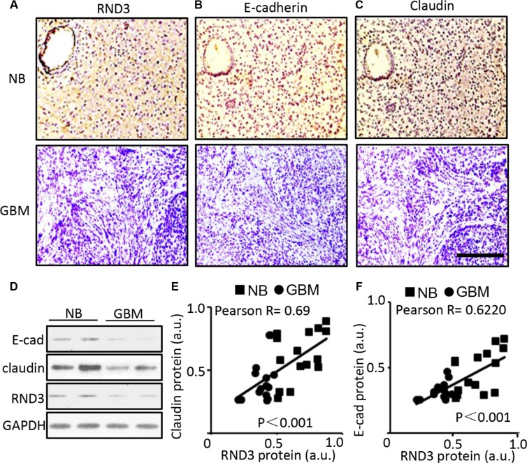
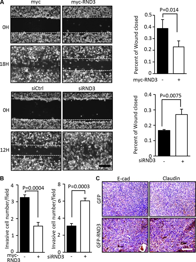
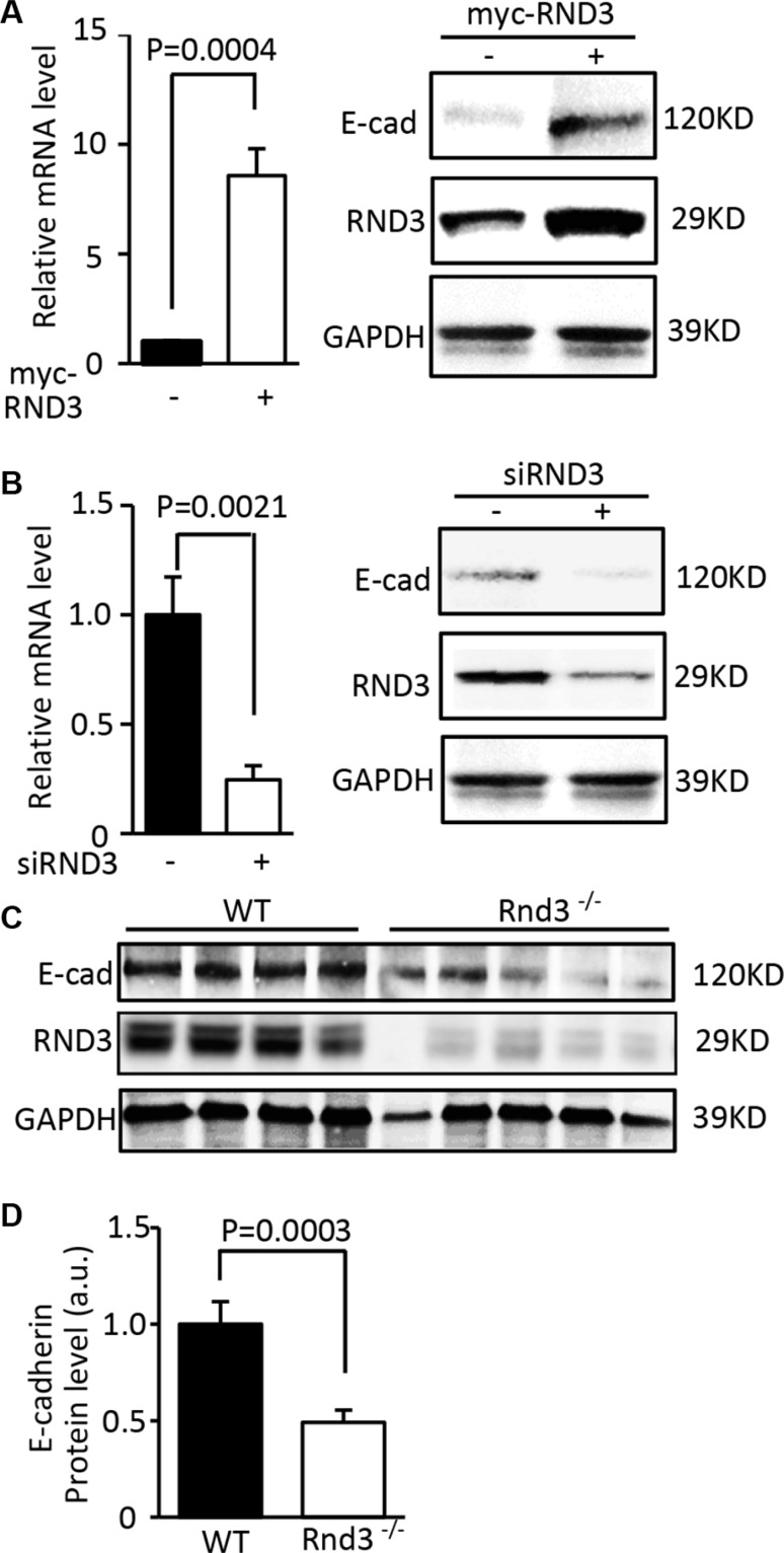
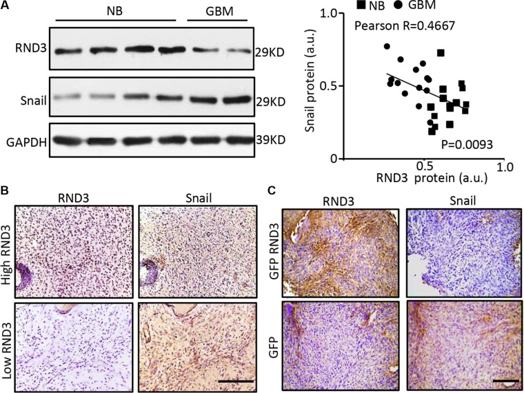
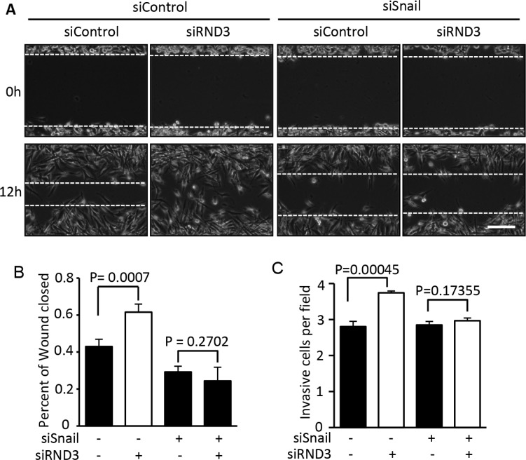
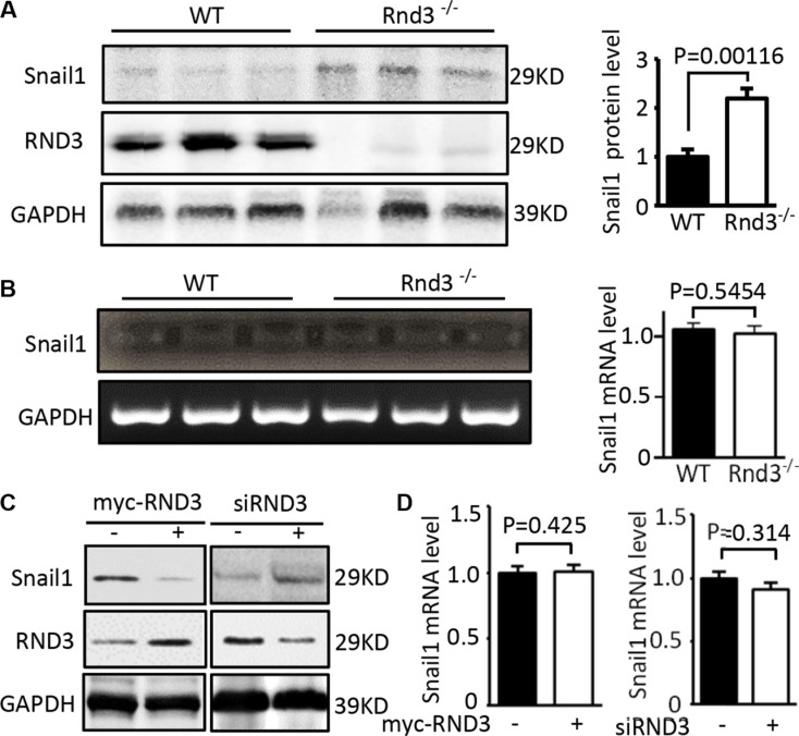
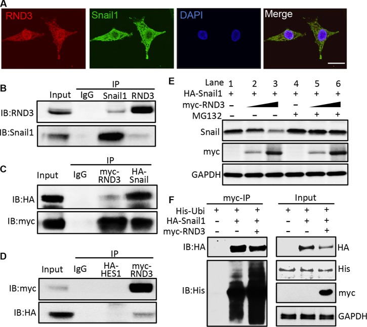
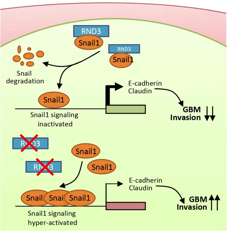
Activation of Snail1 signaling promotes the migration and invasion of multiple tumors, including glioblastoma multiforme (GBM). However, the molecular mechanism that augments Snail1 signaling during GBM cell migration and invasion remains largely unknown. Identification of the factors that regulate Snail1 signaling is critical to block tumor cell migration and invasion. By screening human GBM specimens, we found that the expression levels of small GTPase RND3 positively correlated with the expression levels of E-cadherin and claudin, the glioblastoma migration biomarkers negatively regulated by Snail1. Downregulation of E-cadherin and claudin has been associated with the migration and invasion of GBM cells. We demonstrated that RND3 functioned as an endogenous inhibitor of the Snail-directed transcriptional regulation. RND3 physically interacted with Snail1 protein, enhanced Snail1 ubiquitination, and facilitated the protein degradation. Forced expression of RND3 inhibited Snail1 activity, which in turn blocked glioblastoma cell migration and invasion in vitro in cell culture and in vivo in GBM xenograft mice. In contrast, downregulation of RND3 augmented Snail1 activity, and subsequently decreased E-cadherin expression, eventually promoted glioblastoma cell migration and invasion. The pro-migration induced by RND3 downregulation was attenuated by Snail1 knockdown. The findings partially explain why Snail1 activity is augmented in GBM, and defines a new function of RND3 in GBM cell migration and invasion.

Keywords: RND3; multiform glioblastoma; snail1 signaling.


Conflict of interest statement

CONFLICTS OF INTEREST

There are no conflicts of interest.

Figures

Figure 1
Figure 1. Significant downregulation of E-cadherin, claudin and RND3 protein levels were detected in human GBM tissues
(A–C) Representative immunohisochemical staining (brown) for RND3, E-cadherin, and claudin in human glioblastoma tissues serial sections, respectively. Blue color indicates nuclear staining. (D) Immunoblotting analysis showed the expression levels of RND3 and E-cadherin, claudin in human normal brain (NB) and glioblastoma (GBM). (E–F) Quantification of the immunohistochemical staining for RND3 and E-cadherin and claudin showed a positive correlation of the two protein expression levels in human GBM tissues (n = 15) and brain tissues (n = 15). Statistical analysis of correlation was performed with Pearson's test. Scale bar: 10 μm. a.u.: arbitrary unit.
Figure 2
Figure 2. Forced expression of RND3 inhibited glioblastoma cell migration and invasion
(A–B) U251 cells, a human GBM cell line, were transfected with myc-RND3 or siRND3 to overexpress or knockdown RND3, respectively. Transfected cells were synchronized and cultured in the growth medium to assess the migration and invasion by wound healing and transwell assay, respectively. Migration /invasion of U251 cells were repressed after exogenous RND3 was introduced, while treatment of siRND3 promoted invasion. In right side of panel A, the closure of wound gap was quantified from nine images. In panel B, each experiment was duplicated and motile cells were counted from 20 fields of each experiment (10 field/per duplicates). (C) In vivo E-cadherin and claudin expression in tumors were assessed by immunohistochemical staining (dark red). Tumors generated from nude mice by intracranial implantation of GFP-RND3 cells, a U251 cell lines with GFP-RND3 stable expression, were compared to the tumors derived from the control mice with intracranial implantation of U251 cells expressing GFP. Statistical analysis was performed with the Student's t test in A and paired Student's t test in B. Scale bar represents 100 μm in A and C.
Figure 3
Figure 3. Expression of E-cadherin was closely regulated by RND3
(A) Forced expression of RND 3 upregulated EMT marker E-cadherin transcript (left panel) and protein levels (right panel) in U251 cells. (B) Knockdown of RND 3 resulted in inhibition of E-cadherin transcript (left panel) and protein levels (right panel) in U251 cells. (C) Protein expression level of E-cadherin was significantly decreased in Rnd3-null mouse brain tissues compared to the WT control mice. (D) This downregulation was quantified. Student's t test was used.
Figure 4
Figure 4. RND3 expression levels were inversely correlated with Snail1 expression levels in human and mouse GBM tissues
(A–B) the protein levels of RND3 and Snail1 in human GBM tissues are detected by immunoblotting and immunohistochemical staining (brown). Quantification of the immunoblotting for RND3 and Snail showed an inverse correlation of the two protein expressions in human normal brain (NB) and glioblastoma (GBM) tissues (n = 30) (A, right panel). (C) Significant decrease in snail expression was shown in the GBM xenograft nude mouse brains with intracranial implantation of U251 cells overexpressing GFP-RND3 compared with the control mice with the implantation of U251 cells expressing GFP only. Pearson's test was performed for the correlation analysis. Scale bars represent 100 μm.
Figure 5
Figure 5. Knockdown of Snail1 diminished the promotion of RND3 deficiency-induced GBM cell migration and invasion
The migration and invasiob of GBM cells was assessed by wound healing and transwell assay, respectively. (A–B) The wound healing experiment showed that RND3 deficiency promoted tumor cell migration. This enhancement of the cell migration was completely blocked by Snail knockdown. (C) A similar result was achieved in a transwell assay. Student's t test was used for the statistical analyses. Scale bar represents 100 μm.
Figure 6
Figure 6. RND3 negatively regulated the expression levels of Snail protein but not the transcript
(A) Immunoblot analysis showed that Rnd3 deficiency resulted in increases in the Snail at protein levels in Rnd3 null mouse brain tissues. (B) No changes of Snail1 transcripts were found in the same cohort of Rnd3 null mouse brains. (C) Forced expression of RND3 in U251 cells resulted in a decrease in Snail1 protein level. An opposite result was observed when RND3 was knocked down. (D) The transcript levels of Snail1 remained unchanged under overexpression and downregulation of RND3 conditions. Student's t was used for the statistical analyses.
Figure 7
Figure 7. Rnd3 physically interacted with Snail1 and facilitated Snail1 protein degradation
(A) immunofluorescent staining displayed the colocalization of RND3 (green) and Snail1 (red) expression in U251 cells. (B–D) Co-immunoprecipitation (IP) pull-downs were conducted at both endogenous (B) and exogenous level (2 μg of each plasmid was co-transfected) (C) followed by immunoblotting analysis (IB). The blots demonstrated the interaction of RND3 and Snail1 proteins while no non-specific binding with negative control, HA-HES1 (D). (E) The Snail1 protein levels in the whole cell lysates transfected with the RND3 expression vector were assessed by immunoblotting analysis. Along with the elevations of RND3 protein levels, noticeable decreases in the Snail1 protein levels were observed and this effect was partially attenuated by treatment with a proteasome inhibitor MG-132(10 μM). (F) A representative immunoblotting analysis for ubiquitin showed the increase in the ubiquitination level of Snail1 pull-down complex in the forced expression of RND3 group. Meanwhile, the reduction in Snail1 protein levels was detected in parallel with this enhanced ubiquitination. Scale bar represents 50 μm.
Figure 8
Figure 8. A proposed model outlining the molecular mechanism of the Rnd3 negatively regulating GBM cell migration/invasion through repressing snail signaling
In the presence of RND3, RND3 represses Snail activity by physically interacting with Snail protein and promoting its degradation. In the absence of RND3, the snail signaling is significantly enhanced due to the extra amount of Snail proteins available, which represses E-cadherin and claudin expressions, promoting tumor cell migration and invasion.

References

    1. Ferrari-Amorotti G, Fragliasso V, Esteki R, Prudente Z, Soliera AR, Cattelani S, Manzotti G, Grisendi G, Dominici M, Pieraccioli M, Raschella G, Chiodoni C, Colombo MP, et al. Inhibiting interactions of lysine demethylase LSD1 with snail/slug blocks cancer cell invasion. Cancer research. 2013;73:235–245. - PMC - PubMed
    1. Yang Z, Rayala S, Nguyen D, Vadlamudi RK, Chen S, Kumar R. Pak1 phosphorylation of snail, a master regulator of epithelial-to-mesenchyme transition, modulates snail's subcellular localization and functions. Cancer Res. 2005;65:3179–3184. - PubMed
    1. Cano A, Perez-Moreno MA, Rodrigo I, Locascio A, Blanco MJ, del Barrio MG, Portillo F, Nieto MA. The transcription factor snail controls epithelial-mesenchymal transitions by repressing E-cadherin expression. Nat Cell Biol. 2000;2:76–83. - PubMed
    1. Batlle E, Sancho E, Franci C, Dominguez D, Monfar M, Baulida J, Garcia De Herreros A. The transcription factor snail is a repressor of E-cadherin gene expression in epithelial tumour cells. Nat Cell Biol. 2000;2:84–89. - PubMed
    1. Colvin Wanshura LE, Galvin KE, Ye H, Fernandez-Zapico ME, Wetmore C. Sequential activation of Snail1 and N-Myc modulates sonic hedgehog-induced transformation of neural cells. Cancer Res. 2011;71:5336–5345. - PMC - PubMed

MeSH terms