

Antidermatophytic Action of Resorcinol Derivatives: Ultrastructural Evidence of the Activity of Phenylethyl Resorcinol against Microsporum gypseum
- PMID: 27706019
- PMCID: PMC6274034
- DOI: 10.3390/molecules21101306
Antidermatophytic Action of Resorcinol Derivatives: Ultrastructural Evidence of the Activity of Phenylethyl Resorcinol against Microsporum gypseum
Abstract
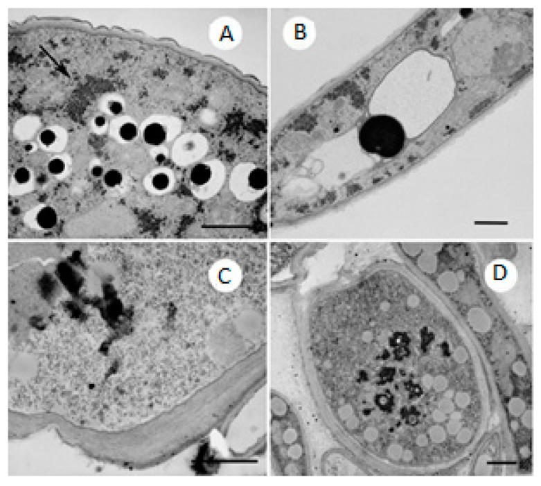
In this work, we evaluated the antidermatophytic activities of three resorcinol derivatives that have a history of use in dermo-cosmetic applications to discover molecules with multiple dermatological activities (i.e., multi-target drugs), thereby reducing the cost and time necessary for new drug development. The antidermatophytic activities of the three skin lighteners were evaluated relative to the known antifungal drug fluconazole on nine dermatophytes responsible for the most common dermatomycoses: Microsporum gypseum, Microsporum canis, Trichophyton violaceum, Arthroderma cajetani, Trichophyton mentagrophytes, Epidermophyton floccosum, Nannizzia gypsea, Trichophyton rubrum and Trichophyton tonsurans. Among the three tested resorcinols, only two showed promising properties, with the ability to inhibit the growth of all tested dermatophytes; additionally, the IC50 values of these two resorcinols against the nine dermatophytes confirmed their good antifungal activity, particularly for phenylethyl resorcinol against M. gypseum. Ultrastructural alterations exhibited by the fungus were observed using scanning electron microscopy and transmission electron microscopy and reflected a dose-dependent response to treatment with the activation of defence and self-preservation strategies.
Keywords: Microsporum gypseum; SEM; TEM; antifungal activity; dermatophytes; resorcinol derivatives.
Conflict of interest statement
The authors declare no conflict of interest.
Figures


Similar articles
-
A Multi-Target Approach toward the Development of Novel Candidates for Antidermatophytic Activity: Ultrastructural Evidence on α-Bisabolol-Treated Microsporum gypseum.Molecules. 2015 Jun 26;20(7):11765-76. doi: 10.3390/molecules200711765. Molecules. 2015. PMID: 26132903 Free PMC article.
-
Antioxidant and antifungal activities of marrubiin, extracts and essential oil from Marrubium vulgare L. against pathogenic dermatophyte strains.J Mycol Med. 2020 Apr;30(1):100927. doi: 10.1016/j.mycmed.2020.100927. Epub 2020 Jan 11. J Mycol Med. 2020. PMID: 31983544
-
Ultrastructural changes on clinical isolates of Trichophyton rubrum, Trichophyton mentagrophytes, and Microsporum gypseum caused by Solanum chrysotrichum saponin SC-2.Planta Med. 2009 Nov;75(14):1517-20. doi: 10.1055/s-0029-1185810. Epub 2009 Jun 23. Planta Med. 2009. PMID: 19551614
-
The Changing Face of Dermatophytic Infections Worldwide.Mycopathologia. 2017 Feb;182(1-2):77-86. doi: 10.1007/s11046-016-0082-8. Epub 2016 Oct 25. Mycopathologia. 2017. PMID: 27783316 Review.
-
Antifungal activity of lichen compounds against dermatophytes: a review.J Appl Microbiol. 2019 Aug;127(2):308-325. doi: 10.1111/jam.14209. Epub 2019 May 8. J Appl Microbiol. 2019. PMID: 30664814 Review.
Cited by
-
Vesicular carriers containing phenylethyl resorcinol for topical delivery system; liposomes, transfersomes and invasomes.Asian J Pharm Sci. 2018 Sep;13(5):472-484. doi: 10.1016/j.ajps.2018.02.004. Epub 2018 Mar 17. Asian J Pharm Sci. 2018. PMID: 32104421 Free PMC article.
-
Antifungal and Anti-Inflammatory Potential of Bupleurum rigidum subsp. paniculatum (Brot.) H.Wolff Essential Oil.Antibiotics (Basel). 2021 May 17;10(5):592. doi: 10.3390/antibiotics10050592. Antibiotics (Basel). 2021. PMID: 34067555 Free PMC article.
-
A rare urinary tract infection of multidrug-resistant Chryseobacterium urinae sp. nov. isolated from a diabetic, non-catheterized patient.Arch Microbiol. 2024 Mar 11;206(4):150. doi: 10.1007/s00203-024-03881-0. Arch Microbiol. 2024. PMID: 38466448
-
Phytofabricated Farsetia aegyptia-derived silver nanoparticles mediate antibacterial and wound-healing activities in diabetic foot infection rat model.Inflammopharmacology. 2025 Jun;33(6):3233-3254. doi: 10.1007/s10787-025-01789-9. Epub 2025 Jun 3. Inflammopharmacology. 2025. PMID: 40461756
-
Stingless bee propolis: a comprehensive review of chemical constituents and health efficacy.Nat Prod Bioprospect. 2025 Sep 4;15(1):61. doi: 10.1007/s13659-025-00545-4. Nat Prod Bioprospect. 2025. PMID: 40906303 Free PMC article. Review.
References
-
- Romagnoli C., Baldisserotto A., Malisardi G., Vicentini C.B., Mares D., Andreotti E., Vertuani S., Manfredini S. A multi-target approach toward the development of novel candidates for antidermatophytic activity: Ultrastructural evidence on α-bisabolol-Treated Microsporum gypseum. Molecules. 2015;20:11765–11776. doi: 10.3390/molecules200711765. - DOI - PMC - PubMed
-
- Martindale W. The Extra Pharmacopoeia. 28th ed. Pharmaceutical Press; London, UK: 1989. pp. 1637–1638.
-
- Birch C.A. Castellani’s paint. Sir Aldo Castellani. (1877–1971) Practitioner. 1974;212:895–896. - PubMed
-
- Adawadkar P.D., Elsohly M.A. Isolation, purification and antimicrobial activity of anacardic acid from Ginko biloba fruits. Fitoterapia. 1981;52:129–135.
MeSH terms
Substances
LinkOut - more resources
Full Text Sources
Other Literature Sources
