Review
. 2016 Oct 3;7(10):81.
doi: 10.3390/genes7100081.

The SaeRS Two-Component System of Staphylococcus aureus

Affiliations
Review

The SaeRS Two-Component System of Staphylococcus aureus

Qian Liu et al. Genes (Basel). .

Abstract

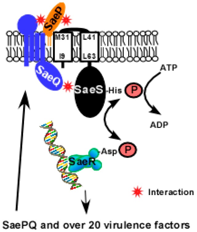
In the Gram-positive pathogenic bacterium Staphylococcus aureus, the SaeRS twocomponent system (TCS) plays a major role in controlling the production of over 20 virulence factors including hemolysins, leukocidins, superantigens, surface proteins, and proteases. The SaeRS TCS is composed of the sensor histidine kinase SaeS, response regulator SaeR, and two auxiliary proteins SaeP and SaeQ. Since its discovery in 1994, the sae locus has been studied extensively, and its contributions to staphylococcal virulence and pathogenesis have been well documented and understood; however, the molecular mechanism by which the SaeRS TCS receives and processes cognate signals is not. In this article, therefore, we review the literature focusing on the signaling mechanism and its interaction with other global regulators.

Keywords: Staphylococcus aureus; Bacterial histidine  kinase; Two‐component system; Virulence factors.


Conflict of interest statement

The authors declare no conflict of interest. The funding sponsors had no role in the design of the study, in the collection, analyses, or interpretation of data, in the writing of the manuscript, and in the decision to publish the results.

Figures

Figure A1
Figure A1
Effect of oxacillin on P1 promoter activity. Staphylococcus aureus USA300 carrying P1-GFP reporter plasmid [42] was grown in 150 µL TSB at 37 °C until mid-exponential growth phase. After addition of oxacillin to the concentration indicated, the culture was further incubated for 2 h at 37 °C, and OD600 and GFP expression were measured (Enspire, Perkin Elmer, Waltham, MA, USA). AU, arbitrary unit.
Figure 1
Figure 1
The sae operon. (A) Organization of the sae operon. Two angled arrows represent the P1 and P3 promoters, respectively. Two vertical lines in the P1 promoter region indicate the SaeR binding sequences (SBSs). The nucleotide sequence of the P3 promoter is shown under saeQ, where N17 = 17 nucleotides. Four transcripts (T1–T4) are indicated by arrows; (B) DNA sequences of select sae target promoters. The SBSs are shown in gray. Transcription start sites are indicated by angled arrows. The promoter sequences are shown in boxes. Pcoa, the promoter of coagulase (coa); Phla, the promoter of α-hemolysin (hla).
Figure 2
Figure 2
The SaeS protein. (A) The domain structure of SaeS. The numbers represent the boundary amino acids. The red star indicates the L18P mutation of SaeS in the strain Newman. N,N-terminus; H, His 131; C,C-terminus; TM, transmembrane helix; HK, histidine kinase; (B) A summary of mutations in the transmembrane domain of SaeS. Increased, Increased basal kinase activity; Decreased, Decreased basal kinase activity; No change, No effect on the basal kinase activity; Insensitive to HNP1, Kinase activity does not respond to HNP1. The positions of amino acids are all predictions.
Figure 3
Figure 3
The consensus sequence of the SaeR binding sequence. It was generated by WebLogo (http://weblogo.berkeley.edu/logo.cgi) using the SaeR binding sequences in Table 1.
Figure 4
Figure 4
A model for SaePQS interaction. Upon exposure to the inducing signal(s), SaeS autophosphorylates the conserved His residue with ATP; then the phosphoryl group is transferred to the conserved Asp residue of SaeR. The phosphorylated SaeR binds to its binding sequence and activates transcription from target promoters including the saeP1 promoter. From saeP1, SaeP and SaeQ are produced and bind to SaeS in the membrane. As a lipoprotein, SaeP is expected to interact with the extracellular linker peptide of SaeS. On the other hand, SaeQ is thought to interact with the cytoplasmic domain of SaeS [22]. The interacting parts of SaePQS are based on predictions.
Figure 5
Figure 5
Organization of SBSs in the sae target gene promoters. Each white triangle represents a half of the SaeR binding sequence. Arrows represent promoter region pointing the direction of transcription. Unless stated otherwise, the distance between each half binding sequence is 6 bp. Numbers are base pairs of the gap between the half-binding sites.

Similar articles

Cited by

References

    1. Lowy F.D. Staphylococcus aureus infections. N. Engl. J. Med. 1998;339:520–532. doi: 10.1056/NEJM199808203390806. - DOI - PubMed
    1. Gordon R.J., Lowy F.D. Pathogenesis of methicillin-resistant Staphylococcus aureus infection. Clin. Infect. Dis. 2008;46:S350–S359. doi: 10.1086/533591. - DOI - PMC - PubMed
    1. Recsei P., Kreiswirth B., O′Reilly M., Schlievert P., Gruss A., Novick R.P. Regulation of exoprotein gene expression in Staphylococcus aureus by agr. Mol. Gen. Genet. 1986;202:58–61. doi: 10.1007/BF00330517. - DOI - PubMed
    1. Xiong Y.Q., Willard J., Yeaman M.R., Cheung A.L., Bayer A.S. Regulation of Staphylococcus aureus α-toxin gene (hla) expression by agr, sarA, and sae in vitro and in experimental infective endocarditis. J. Infect. Dis. 2006;194:1267–1275. doi: 10.1086/508210. - DOI - PubMed
    1. Cheung G.Y., Joo H.S., Chatterjee S.S., Otto M. Phenol-soluble modulins—Critical determinants of staphylococcal virulence. FEMS Microbiol. Rev. 2014;38:698–719. doi: 10.1111/1574-6976.12057. - DOI - PMC - PubMed

LinkOut - more resources